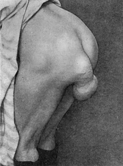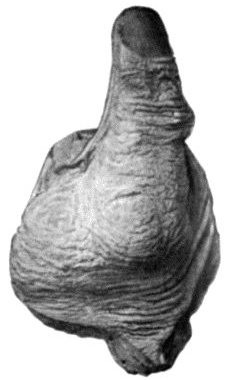
Fig. 45.—Subcutaneous Lipoma showing lobulation.
Lipoma.—A lipoma is composed of fat resembling that normally present in the body. The commonest variety is the subcutaneous lipoma, which grows from the subcutaneous fat, and forms a soft, irregularly lobulated tumour (Fig. 45). The fat is arranged in lobules separated by connective-tissue septa, which are continuous with the capsule surrounding the tumour and with the overlying skin, which becomes dimpled or puckered when an attempt is made to pinch it up. As the fat is almost fluid at the body temperature, fluctuation can usually be detected. These tumours vary greatly in size, occur at all ages, grow slowly, and, while generally solitary, are sometimes multiple. They are most commonly met with on the shoulder, buttock, or back. In certain situations, such as the thigh and perineum, they tend to become pedunculated (Fig. 46).
A fatty tumour is to be diagnosed from a cold abscess and from a cyst. The distinguishing features of the lipoma are the tacking down and dimpling of the overlying skin, the lobulation of the tumour, which is recognised when it is pressed upon with the flat of the hand, and, more reliable than either of these, the mobility, the tumour slipping away when pressed upon at its margin.
The prognosis is more favourable than in any other tumour as it never changes its characters; the only reasons for its removal by operation are its unsightliness and its probable increase in size in the course of years. The operation consists in dividing the skin and capsule over the tumour and shelling it out. Care must be taken that none of the outlying lobules are left behind. If the overlying skin is damaged or closely adherent, it should be removed along with the tumour.
Multiple subcutaneous lipomas are frequently symmetrical, and in a certain group of cases, met with chiefly in women, pain is a prominent symptom, hence the term adiposis dolorosa (Dercum). These multiple tumours show little or no tendency to increase in size, and the pain which attends their development does not persist.
In the neck, axilla, and pubes a diffuse overgrowth of the subcutaneous fat is sometimes met with, forming symmetrical tumour-like masses, known as diffuse lipoma. As this is not, strictly speaking, a tumour, the term diffuse lipomatosis is to be preferred. A similar condition was described by Jonathan Hutchinson as being met with in the domestic animals. If causing disfigurement, the mass of fat may be removed by operation.
Lipoma in other Situations.—The periosteal lipoma is usually congenital, and is most often met with in the hand; it forms a projecting lobulated tumour, which, when situated in the palm, resembles an angioma or a lymphangioma. The subserous lipoma arises from the extra-peritoneal fat in the posterior abdominal wall, in which case it tends to grow forwards between the layers of the mesentery and to give rise to an abdominal tumour; or it may grow from the extra-peritoneal fat in the anterior abdominal wall and protrude from one of the hernial openings or through an abnormal opening in the parietes, constituting a fatty hernia. A subsynovial lipoma grows from the fat surrounding the synovial membrane of a joint, and projects into its interior, giving rise to the symptoms of loose body. Lipomas are also met with growing from the adipose connective tissue between or in the substance of muscles, and, when situated beneath the deep fascia, such as the fascia lata of the thigh, the characteristic signs are obscured and a differential diagnosis is difficult. It may be differentiated from a cold abscess by puncture with an exploring needle.
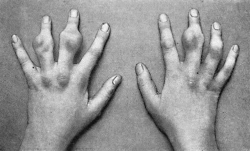
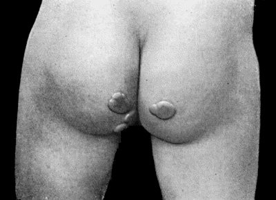
Fig. 48.—Zanthoma of Hands in a girl æt. 14, showing multiple subcutaneous tumours (cf. Fig. 49).
(Sir H. J. Stiles' case.)
Zanthoma is a rare but interesting form of tumour, composed of a fibrous and fatty tissue, containing a granular orange-yellow pigment, resembling that of the corpus luteum. It originates in the corium and presents two clinical varieties. In the first of these, it occurs in the form of raised yellow patches, usually in the skin of the eyelids of persons after middle life, and in many instances is associated with chronic jaundice; the patches are often symmetrical, and as they increase in size they tend to fuse with another.
The second form occurs in children and adolescents; it may affect several generations of the same family, and is often multiple, there being a combination of thickened yellow patches of skin and projecting tumours, some of which may attain a considerable size (Figs. 48 and 49). On section, the tumour tissue presents a brilliant orange or saffron colour.
There is no indication for removing the tumours unless for the deformity which they cause; exposure to the X-rays is to be preferred to operation.
Chondroma.—A chondroma is mainly composed of cartilage. Processes of vascular connective tissue pass in between the nodules of cartilage composing the tumour from the fibrous capsule which surrounds it. On section it is of a greyish-blue colour and semi-translucent. The tumour is firm and elastic in consistence, but certain portions may be densely hard from calcification or ossification, while other portions may be soft and fluctuating as a result of myxomatous degeneration and liquefaction. These tumours grow slowly and painlessly, and may surround nerves and arteries without injuring them. They may cause a deep hollow in the bone from which they originate. All intermediate forms between the innocent chondroma and the malignant chondro-sarcoma are met with. Chondroma may occur in a multiple form, especially in relation to the phalanges and metacarpal bones. When growing in the interior of a bone it causes a spindle-shaped enlargement of the shaft, which in the case of a phalanx or metacarpal bone may resemble the dactylitis resulting from tubercle or syphilis. A chondroma appears as a clear area in a skiagram.
A skiagram of a bone in which there is a chondroma shows a clear rounded area in the position of the tumour, which must be differentiated from similar clear areas due to other kinds of tumour, especially the myeloma; when it has undergone calcification or ossification, it gives a shadow as dark as bone.
Treatment.—In view of the unstable quality of the chondroma, especially of its liability to become malignant, it should be removed as soon as it is recognised. In those projecting from the surface of a bone, both the tumour and its capsule should be removed. If in the interior, a sufficient amount of the cortex should be removed to allow of the tumour being scraped out, and care must be taken that no nodules of cartilage are left behind. In multiple chondromas of the hand, when the fingers are crippled and useless, exposure to the X-rays should be given a trial, and in extreme cases the question of amputation may have to be considered. When a cartilaginous tumour takes on active growth, it must be treated as malignant.
The chondromas that are met with at the ends of the long bones in children and young adults form a group by themselves. They are usually related to the epiphysial cartilage, and it was suggested by Virchow that they take origin from islands of cartilage which have not been used up in the process of ossification. They are believed to occur more frequently in those who have suffered from rickets. They have no malignant tendencies and tend to undergo ossification concurrently with the epiphysial cartilage from which they take origin, and constitute what are known as cartilaginous exostoses. These are sometimes met with in a multiple form, and may occur in several generations of the same family. They are considered in greater detail in the chapter dealing with tumours of bone.
Minute nodules of cartilage sometimes form in the synovial membrane of joints and lining of tendon sheaths and bursæ: they tend to become detached from the membrane and constitute loose bodies; they also undergo a variable amount of calcification and ossification, so as to be visible in skiagrams. They are further considered with loose bodies in joints.
Cartilaginous tumours in the parotid, submaxillary gland, and testicle belong to a class of “mixed tumours” that will be referred to later.
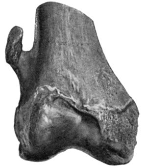
Osteoma.—The true osteoma is composed of bony tissue, and originates from the skeleton. Two varieties are recognised—the spongy or cancellous, and the ivory or compact. The spongy or cancellous osteoma is really an ossified chondroma, and is met with at the ends of the long bones (Fig. 52). From the fact that it projects from the surface of the bone it is often spoken of as an exostosis. It grows slowly, and rarely causes any discomfort unless it presses upon a nerve-trunk or upon a bursa which has developed over it. The Röntgen rays show a dark shadow corresponding to the ossified portion of the tumour, and continuous with that of the bone from which it is growing (Fig. 138). Operative interference is only indicated when the tumour is giving rise to inconvenience. It is then removed, its base or neck being divided by means of the chisel. The multiple variety of osteoma is considered with the diseases of bone.
The bony outgrowth from the terminal phalanx of the great toe—known as the subungual exostosis—is described and figured on p. 404. Bony projections or “spurs” sometimes occur on the under surface of the calcaneus, and, projecting downwards and forwards from the greater process, cause pain on putting the heel to the ground.
The ivory or compact osteoma is composed of dense bone, and usually grows from the skull. It is generally sessile and solitary, and may grow into the interior of the skull, into the frontal sinus, into the cavity of the orbit or nose, or may fill up the external auditory meatus, causing most unsightly deformity and interference with sight, breathing, and hearing.
Bony formations occur in muscles and tendons, especially at their points of attachment to the skeleton, and are known as false exostoses; they are described with the diseases of muscles.
Odontoma.—An odontoma is composed of dental tissues in varying proportions and different degrees of development, arising from tooth-germs or from teeth still in process of growth (Bland Sutton). Odontomas resemble teeth in so far that during their development they remain hidden below the mucous membrane and give no evidence of their existence. There then succeeds, usually between the twentieth and twenty-fifth years, an eruptive stage, which is often attended with suppuration, and this may be the means of drawing attention to the tumour. Following Bland Sutton, several varieties of odontoma may be distinguished according to the part of the tooth-germ concerned in their formation.
The epithelial odontoma is derived from persistent portions of the epithelium of the enamel organ, and constitutes a multilocular cystic tumour which is chiefly met with in the mandible. The cystic spaces of the tumour contain a brownish glairy fluid. These tumours have been described by Eve under the name of multilocular cystic epithelial tumours of the jaw.
The follicular odontoma, also known as a dentigerous cyst, is derived from the distension of a tooth follicle. It constitutes a cyst containing a viscid fluid, and an imperfectly formed tooth is often found embedded in its wall. The cyst usually forms in relation to one of the permanent molars, and may attain considerable dimensions.
The fibrous odontoma is the result of an overgrowth of fibrous tissue surrounding the tooth sac, which encapsulates the tooth and prevents its eruption. The thickened tooth sac is usually mistaken for a fibrous tumour, until, after removal, the tooth is recognised in its interior.
Composite Odontoma.—This is a convenient term to apply to certain hard dental tumours which are met with in the jaws, and consist of enamel, dentine, and cement. The tumour is to be regarded as being derived from an abnormal growth of all the elements of a tooth germ, or of two or more tooth germs, indiscriminately fused with one another. It may appear in childhood, and form a smooth unyielding tumour, often of considerable size, replacing the corresponding permanent tooth. It may cause a purulent discharge, and in some cases it has been extruded after sloughing of the overlying soft parts. Many examples of this variety of odontoma, growing in the nasal cavity or in the maxillary sinus, have been erroneously regarded as osteomas even after removal.
On section, the tumour is usually laminated, and is seen to consist mainly of dentine with a partial covering of enamel and cement.
Diagnosis.—Odontomas are often only diagnosed after removal. When attended with suppuration, the condition has been mistaken for disease of the jaw. Fibrous odontomas have been mistaken for sarcoma, and portions of the maxilla removed unnecessarily. Any circumscribed tumour of the jaw, particularly when met with in a young adult, should suggest the possibility of an odontoma. Skiagrams often give useful information both for diagnosis and for treatment.
Treatment.—The solid varieties of odontoma can usually be shelled out after dividing the overlying soft parts. In the follicular variety, it is usually sufficient to excise a portion of the wall, scrape out the interior, and remove any tooth that may be present. The cavity is then packed and allowed to heal from the bottom.
Fibroma.—A fibroma is a tumour composed of fibrous connective tissue. A distinction may be made between the soft fibroma, which is comparatively rich in cells and blood vessels, and in which the fibres are arranged loosely; and the hard fibroma, which is composed of closely packed bundles of fibres often arranged in a concentric fashion around the blood vessels. The cut surface of the soft fibroma presents a pinkish-white, fleshy appearance, resembling the slowly growing forms of sarcoma; that of a hard fibroma presents a dry, glistening appearance, aptly compared to watered silk. The soft variety grows much more rapidly than the hard. In certain fibromas—in those, for example, which grow from the periosteum of the base of the skull and project into the naso-pharynx—the blood vessels are dilated into sinuses and have no proper sheaths; they therefore tend to remain open when divided, and to bleed excessively. Transition forms between soft fibroma and sarcoma are met with, so that in operating for their removal it is safer to take away the capsule along with the tumour, and the patient should be kept under observation in view of the risk of recurrence.
The skin—especially the skin of the buttock—is one of the favourite seats of fibroma, and it may occur in a multiple form. It is met with also in the subcutaneous and intermuscular cellular tissue, and in the abdominal wall, where it sometimes attains considerable dimensions. Various forms of fibroma are met with in the mamma and are described with diseases of that organ. The fibrous overgrowths in the skin, known as keloid and molluscum fibrosum, and those met with in the sheaths of nerves, are described elsewhere. Fibroid tumours of the uterus are described with myoma.
Diffuse fibroma or Fibromatosis, analogous to lipomatosis, is met with in the connective tissue of the skin and sheaths of nerves, and constitutes one form of neuro-fibromatosis; a similar change is also met with in the stomach and colon.
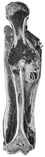
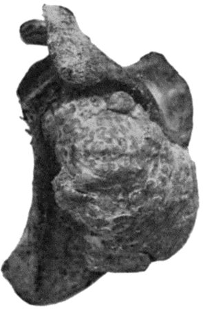
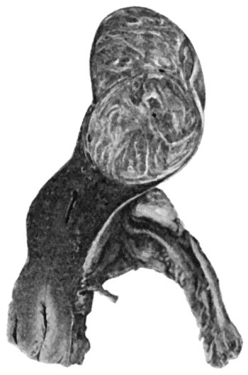
Fig. 53.—Myeloma of Shaft of Humerus, causing pathological fracture. (Mr. J. W. Struthers' case.)
(The unusual site of the tumour is to be noted.)
Myxoma.—A myxoma is composed of tissue of a soft gelatinous, semifluid consistence. The pure myxoma is extremely rare, and clinically resembles the lipoma. Myxomatous tissue is, however, frequently found in other connective-tissue tumours as a result of degeneration, for example, in cartilaginous tumours and in sarcomas. Myxomatous tissue is also a prominent constituent of the “innocent parotid tumour.” Mucous polypus of the nose, which is often described as a myxoma, is merely a pendulous process of œdematous mucous membrane.
Myeloma.—A myeloma is composed of large multinuclear giant cells surrounded by round and spindle cells. The cut surface of the tumour presents a deep red or maroon colour. While occasionally met with in tendon sheaths and bursæ, and is then of an orange-yellow colour, the myeloma occurs most frequently in the cancellous tissue at the ends of the long bones, its favourite site being the upper end of the tibia. Although formerly classified as a sarcoma, it is the exception for it to present malignant features, and it can usually be extirpated by local measures without fear of recurrence. The diagnosis, X-ray appearances, and the method of removal are considered with the diseases of bone. Sometimes the myeloma is met with in multiple form in the skeleton, in association with an unusual form of protein in the urine (Bence Jones).
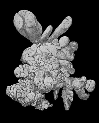
Myoma.—A myoma is composed of non-striped muscle fibres. A pure myoma is very rare, and is met with in organs possessed of non-striped muscle, such as the stomach, intestine, urinary bladder, and prostate. In the uterus, which is the most common situation, these tumours contain a considerable admixture of fibrous tissue, and are known as fibroids or fibro-myomas. They present on section a fasciculated appearance, which may resemble that of a section of balls of cotton (Fig. 54). They are encapsulated and vascular, frequently attain a large size, and may be single or multiple. While they may occasion neither inconvenience nor suffering, they frequently give rise to profuse hæmorrhage from the uterus, and may cause serious symptoms by pressing injuriously on the ureters or the intestine, or by complicating pregnancy and parturition.
The Rhabdomyoma is an extremely rare form of tumour, met with in the kidney, uterus, and testicle. It contains striped muscle fibres, and is supposed to originate from a residue of muscular tissue which has become sequestrated during development.
Glioma.—A glioma is a tumour composed of neuroglia. It is met with exclusively in the central nervous system, retina, and optic nerve. It is a slowly growing, soft, ill-defined tumour, which displaces the adjacent nerve centres and nerve tracts, and is liable to become the seat of hæmorrhage and thus to give rise to pressure symptoms resembling apoplexy. The glioma of the retina tends to grow into the vitreous humour and to perforate the globe. It is usually of the nature of a glio-sarcoma and is highly malignant.
Endotheliomas take origin from the endothelium of lymph vessels and blood vessels, and serous cavities. They show great variation in type, partly because of the number of different kinds of endothelium from which they are derived, and partly because the new connective tissue which is formed is liable to undergo transformation into other tissues. They may be soft or hard, solid or cystic, diffuse or circumscribed; they grow very slowly, and are almost always innocent, although recurrence has been occasionally observed. Cases of multiple endotheliomata of the skin have recently been described by Wise.
Angioma, lymphangioma, and neuroma are described with the disease of the individual tissues.