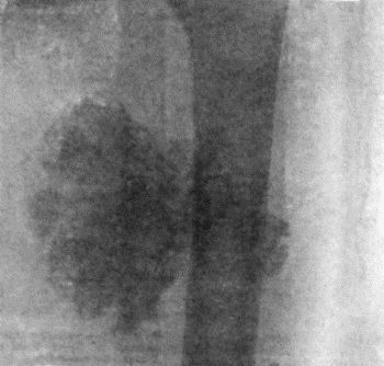
Fig. 138.—Radiogram of Right Knee showing Multiple Exostoses.
New growths which originate in the skeleton are spoken of as primary tumours; those which invade the bones, either by metastasis from other parts of the body or by spread from adjacent tissues, as secondary. A tumour of bone may grow from the cellular elements of the periosteum, the marrow, or the epiphysial cartilage.
Primary tumours are of the connective-tissue type, and are usually solitary, although certain forms, such as the chondroma, may be multiple from the outset.
Periosteal tumours are at first situated on one side of the bone, but as they grow they tend to surround it completely. Innocent periosteal tumours retain the outer fibrous layer as a capsule. Malignant tumours tend to perforate the periosteal capsule and invade the soft parts.
Central or medullary tumours as they increase in size replace the surrounding bone, and simultaneously new bone is formed on the surface; as this is in its turn absorbed, further bone is formed beneath the periosteum, so that in time the bone is increased in girth, and is said to be “expanded” by the growth in its interior.
Primary Tumours—Osteoma.—When the tumour projects from the surface of a bone it is called an exostosis. When growing from bones developed in membrane, such as the flat bones of the skull, it is usually dense like ivory, and the term ivory exostosis is employed. When derived from hyaline cartilage—for example, at the ends of the long bones—it is known as a cartilaginous exostosis. This is invested with a cap of cartilage from which it continues to grow until the skeleton attains maturity.
An exostosis forms a rounded or mushroom-shaped tumour of limited size, which may be either sessile or pedunculated, and its surface is smooth or nodulated (Figs. 138 and 139). A cartilaginous exostosis in the vicinity of a joint may be invested with a synovial sac or bursa—the so-called exostosis bursata. The bursa may be derived from the synovial membrane of the adjacent joint with which its cavity sometimes communicates, or it may be of adventitious origin; when it is the seat of bursitis and becomes distended with fluid, it may mask the underlying exostosis, which then requires a radiogram for its demonstration.
Clinically, the osteoma forms a hard, indolent tumour attached to a bone. The symptoms to which it gives rise depend on its situation. In the vicinity of a joint, it may interfere with movement; on the medial side of the knee it may incapacitate the patient from riding. When growing from the dorsum of the terminal phalanx of the great toe—subungual exostosis—it displaces the nail, and may project through its matrix at the point of the toe, while the soft parts over it may be ulcerated from pressure (Fig. 107). It incapacitates the patient from wearing a boot. When it presses on a nerve-trunk it causes pains and cramps. In the orbit it displaces the eyeball; in the nasal fossæ and in the external auditory meatus it causes obstruction, which may be attended with ulceration and discharge. In the skull it may project from the outer table, forming a smooth rounded swelling, or it may project from the inner table and press upon the brain.
The diagnosis is to be made by the slow growth of the tumour, its hardness, and by the shadow which it presents with the X-rays (Fig. 138).
An osteoma which does not cause symptoms may be left alone, as it ceases to grow when the skeleton is mature and has no tendency to change its benign character. If causing symptoms, it is removed by dividing the neck or base of the tumour with a chisel, care being taken to remove the whole of the overlying cartilage. The dense varieties met with in the bones of the skull present greater difficulties; if it is necessary to remove them, the base or neck of the tumour is perforated in many directions with highly tempered drills rotated by some form of engine, and the division is completed with the chisel.
Multiple Exostoses.—This disease, which, by custom, is still placed in the category of tumours, is to be regarded as a disorder of growth, dating from intra-uterine life and probably due to a disturbance in the function of the glands of internal secretion, the thyreoid being the one which is most likely to be at fault (Arthur Keith). The disorder of growth is confined to those elements of the skeleton where a core of bone formed in cartilage comes to be encased in a sheath of bone formed beneath the periosteum. To indicate this abnormality the name diaphysial aclasis has been employed by Arthur Keith at the suggestion of Morley Roberts.
Bones formed entirely in cartilage are exempt, namely, the tarsal and carpal bones, the epiphyses of the long bones, the sternum, and the bodies of the vertebræ. Bones formed entirely in membrane, that is, those of the face and of the cranial vault, are also exempt. The disorder mainly affects the ossifying junctions of the long bones of the extremities, the vertebral border of the scapula, and the cristal border of the ilium.
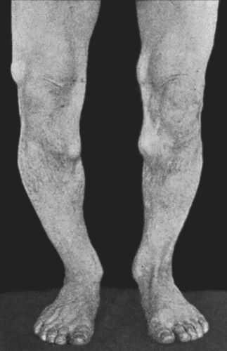
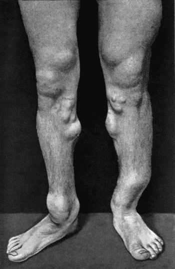
Clinically the disease is attended with the gradual and painless development during childhood or adolescence of a number of tumours or irregular projections of bone, at the ends of the long bones, the vertebral border of the scapula, and the cristal border of the ilium. They exhibit a rough symmetry; they rarely attain any size; and they usually cease growing when the skeleton attains maturity—the conversion of cartilage into bone being then completed. While they originate from the ossifying junctions of the long bones, they tend, as the shaft increases in length, to project from the surface of the bone at some distance from the ossifying junction and to “point” away from it. They may cause symptoms by “locking” the adjacent joint or by pressing upon nerve-trunks or blood vessels.
In a considerable proportion of cases, the disturbance of growth is further manifested by dwarfing of the long bones; these are not only deficient in length but are sometimes also curved and misshapen, which accounts for the condition being occasionally confused with the disturbances of growth resulting from rickets. In about one-third of the recorded cases there is a dislocation of the head of the radius on one or on both sides, a result of unequal growth between the bones of the forearm.
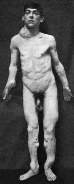
Fig. 140.—Multiple Cartilaginous Exostoses in a man æt. 27. The scapular tumour projecting above the right clavicle has taken on active growth and pressed injuriously on the cords of the brachial plexus.
In early adult life, one of the tumours, instead of undergoing ossification, may take on active growth and exhibit the features of a chondro-sarcoma, pressing injuriously upon adjacent structures (Fig. 140) and giving rise later to metastases in the lungs.
The X-ray appearances of the bones affected are of a striking character; apart from the outgrowths of bone or “tumours” there is evident a widespread alteration in the internal architecture of the bones, which suggests analogies with other disturbances of ossification such as achondroplasia and osteomyelitis fibrosa. The condition is one that runs in families, sometimes through several generations; we have more than once seen a father and son together in the hospital waiting-room.
As regards treatment, there is no indication for surgical interference except when one or other tumour is a source of disability as by pressing upon a nerve-trunk or by locking a joint, in which case it is easily removed by chiselling through its neck.
Diffuse Osteoma, Leontiasis Ossea.—This rare affection was described by Virchow, and named leontiasis ossea because of the disfigurement to which it gives rise. It usually commences in adolescence as a diffuse overgrowth first of one and then of both maxillæ; these bones are enlarged in all directions and project on the face, and the nasal fossæ and the maxillary and frontal sinuses become filled up with bone, which encroaches also on the orbital cavities. In addition to the hideous deformity, the patient suffers from blocking of the nose, loss of smell, and protrusion of the eyes, sometimes followed by loss of sight. The condition is liable to spread to the zygomatic and frontal bones, the vault of the skull, and to the mandible. The base of the skull is not affected. The disease is of slow progress and may become arrested; life may be prolonged for many years, or may be terminated by brain complications or by intercurrent affections. In certain cases it is possible to remove some of the more disfiguring of the bony masses.
A less aggressive form, confined to the maxilla on one side, is sometimes met with, and, in a case of this variety under our own observation, the disfigurement, which was the only subject of complaint, was removed, after reflecting the soft parts, by paring away the excess of bone; this is easily done as the bone is spongy, and at an early stage, imperfectly calcified.
A remarkable form of unilateral hypertrophy and diffuse osteoma of the skull, following the distribution of the fifth nerve, has seen described by Jonathan Hutchinson and Alexis Thomson.
Chondroma.—Cartilaginous tumours, apart from those giving rise to multiple exostoses, grow from the long bones and from the scapula, ilium, ribs, or jaws. They usually project from the surface of the bone, and may attain an enormous size; sometimes they grow in the interior of a bone, the so-called enchondroma.
The hyaline cartilage composing the tumour frequently undergoes myxomatous degeneration, resulting in the formation of a glairy, semi-fluid jelly, and if this change takes place throughout the tumour it comes to resemble a cyst. On the other hand, the cartilage may undergo calcification or ossification. The most important transition of all is that into sarcoma, the so-called malignant chondroma or chondro-sarcoma, which is associated with rapid increase in size, and parts of the tumour may be carried off in the blood-stream and give rise to secondary growths, especially in the lungs.
Cases have been met with in which certain parts of the skeleton—only those developed in cartilage—were so uniformly permeated with cartilage that the condition has been described as a “chondromatosis” and is regarded as dating from an early period of fœtal life. Unlike the condition known as multiple cartilaginous exostoses, it is a malignant disease.
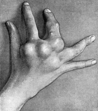
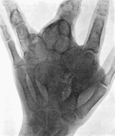
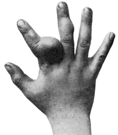
The chondroma is met with as a slowly growing tumour which is specially common in the bones of the hand, often in a multiple form (Figs. 142 and 144). The surface is smooth or lobulated, and in consistence the tumour may be dense and elastic like normal cartilage, or may present areas of softening, or of bony hardness. The skin moves freely over it, except in relation to the bones of the fingers, where it may become adherent and ulcerate, simulating the appearance of a malignant tumour. Large tumours growing from the bones of the extremities may implicate the main vessels and nerves, either surrounding them or pressing on them.
Portions of a chondroma, which have undergone calcification or ossification, throw a dark shadow with the X-rays; unaltered cartilage and myxomatous tissue appear as clear areas.
Treatment.—It is necessary to remove the whole tumour, and in chondromas growing from the surface of the bone, especially if they are pedunculated, this is comparatively easy. When a bone, such as the scapula or mandible, is involved, it is better to excise the bone, or at least the part of it which bears the tumour. In the case of central tumours the shell of bone is removed over an area sufficient to allow of the enucleation of the tumour, or the affected portion of bone is resected. Should there be evidence of malignancy, such as increased rate of growth, a tube of radium should be inserted, and in advanced cases with destruction of tissue, amputation may be called for.
In multiple chondromas of the hand in young subjects, it was formerly the custom to amputate the limb; an attempt should be made to avoid this by shelling out the larger tumours individually, and persevering with the application of the X-rays or of radium to inhibit the growth of the smaller ones.
Chondromas springing from the pelvic bones usually arise in the region of the sacro-iliac joint; they project into the pelvis and press on the bladder and rectum, and on the sciatic and obturator nerves; sometimes also on the iliac veins, causing œdema of the legs. They are liable to take on malignant characters, and rarely lend themselves to complete removal by operation.
Fibroma is met with chiefly as a periosteal growth in relation to the mouth and pharynx, the simple epulis of the alveolar margin and the naso-pharyngeal polypus being the most common examples. We have met with a fibroma in the interior of the lower end of the femur of an adult, causing expansion of the bone with decided increase in girth and liability to pathological fracture; it is possible that this represents the cured stage of osteomyelitis fibrosa.
Myxoma, lipoma, and angioma of bone are all rare.
Myeloma.—The myeloid tumour, which is sometimes classified with the sarcomas, contains as its chief elements large giant cells, like those normally present in the marrow. On section these tumours present a brownish-red or chocolate colour, and, being highly vascular, are liable to hæmorrhages, and therefore also to pigmentation, and to the formation of blood cysts. Sometimes the arterial vessels are so dilated as to impart to the tumour an aneurysmal pulsation and bruit. The enlargement or “expansion” of the bone results in the cortex being represented by a thin shell of bone, which may crackle on pressure—parchment or egg-shell crackling.
The myeloma is most often met with between the ages of twenty-five and forty in the upper end of the tibia or lower end of the femur. It grows slowly and causes little pain, and may long escape recognition unless an examination is made with the X-rays. Although these tumours have been known to give rise to metastases, they are, as a rule, innocent and are to be treated as such. When located in the shaft of a long bone, pathological fracture is liable to occur.
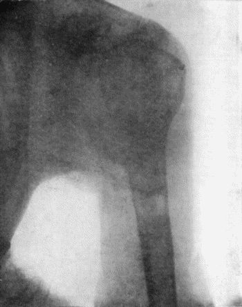
Diagnosis and X-ray Appearances of Myeloma.—The early diagnosis of myeloma is made with the aid of the X-rays: the typical appearance is that of a rounded or oval clear area bounded by a shell of bone of diminishing thickness (Fig. 145). The inflammatory lesions at the ends of the long bones—tubercle, syphilitic gumma, and Brodie's abscess, that resemble myeloma, are all attended with the formation of new bone in greater or lesser amount. The myeloma is also to be diagnosed from chondroma, from sarcoma, and from osteomyelitis fibrosa cystica.
Treatment.—In early cases the cortex is opened up to give free access to the tumour tissue, which is scraped out with the spoon. Bloodgood advises the use of Esmarch's tourniquet, and that the curetting be followed by painting with pure carbolic acid and then rinsing with alcohol; a rod of bone is inserted to fill the gap. In advanced cases the segment of bone is resected and a portion of the tibia or fibula from the other limb inserted into the gap; a tube of radium should also be introduced.
The coexistence of diffuse myelomatosis of the skeleton and albumosuria (Bence-Jones) is referred to on p. 474. Myeloma occurs in the jaws, taking origin in the marrow or from the periosteum of the alveolar process, and is described elsewhere.
Sarcoma and endothelioma are the commonest tumours of bone, and present wide variations in structure and in clinical features. Structurally, two main groups may be differentiated: (1) the soft, rapidly growing cellular tumours, and (2) those containing fully formed fibrous tissue, cartilage, or bone.
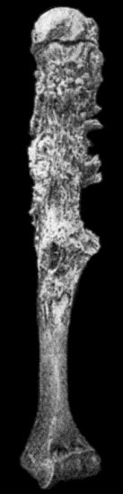
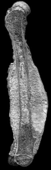
Fig. 147.—Periosteal Sarcoma of Humerus, after maceration.
(Anatomical Museum, University of Edinburgh.)
(1) The soft cellular tumours are composed mainly of spindle or round cells; they grow from the marrow of the spongy ends or from the periosteum of the long bones, the diploë of the skull, the pelvis, vertebræ, and jaws. As they grow they may cause little alteration in the contour of the bone, but they eat away its framework and replace it, so that the continuity of the bone is maintained only by tumour tissue, and pathological fracture is a frequent result. The small round-celled sarcomas are among the most malignant tumours of bone, growing with great rapidity, and at an early stage giving rise to secondary growths.
(2) The second group includes the fibro-, osteo-, and chondro-sarcomas, and combinations of these; in all of them fully formed tissues or attempts at fully formed tissues predominate over the cellular elements. They grow chiefly from the deeper layer of the periosteum, and at first form a projection on the surface, but later tend to surround the bone (Fig. 150), and to invade its interior, filling up the marrow spaces with a white, bone-like substance; in the flat bones of the skull they may traverse the diploë and erupt on the inner table. The tumour tissue next the shaft consists of a dense, white, homogeneous material, from which there radiate into the softer parts of the tumour, spicules, needles, and plates, often exhibiting a fan-like arrangement (Fig. 151). The peripheral portion consists of soft sarcomatous tissue, which invades the overlying soft parts. The articular cartilage long resists destruction. The ossifying sarcoma is met with most often in the femur and tibia, less frequently in the humerus, skull, pelvis, and jaws. In the long bones it may grow from the shaft, while the chondro-sarcoma more often originates at the extremities. Sometimes they are multiple, several tumours appearing simultaneously or one after another. Secondary growths are met with chiefly in the lungs, metastasis taking place by way of the veins.
Clinical Features.—Sarcoma is usually met with before the age of thirty, and is comparatively common in children. Males suffer oftener than females, in the proportion of two to one.
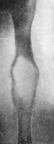
In periosteal sarcoma the presence of a swelling is usually the first symptom; the tumour is fusiform, firm, and regular in outline, and when it occurs near the end of a long bone the limb frequently assumes a characteristic “leg of mutton” shape (Fig. 146). The surface may be uniform or bossed, the consistence varies at different parts, and the swelling gradually tapers off along the shaft. On firm pressure, fine crepitation may be felt from crushing of the delicate framework of new bone.
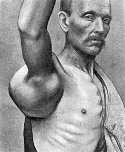
Fig. 148.—Chondro-Sarcoma of Scapula in a man æt. 63; removal of the scapula was followed two years later by metastases and death.
In central sarcoma pain is the first symptom, and it is usually constant, dull, and aching; is not obviously increased by use of the limb, but is often worse at night. Swelling occurs late, and is due to expansion of the bone; it is fusiform or globular, and is at first densely hard, but in time there may be parchment-like or egg-shell crackling from yielding of the thin shell. The swelling may pulsate, and a bruit may be heard over it. In advanced cases it may be impossible to differentiate between a periosteal and a central tumour, either clinically or after the specimen has been laid open.
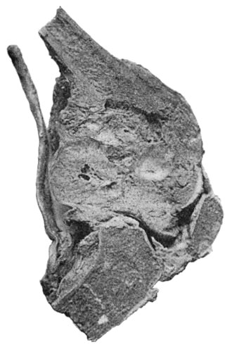
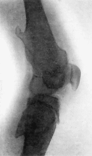
Fig. 149.—Central Sarcoma of Lower End of Femur, invading the knee-joint.
(Museum of Royal College of Surgeons, Edinburgh.)
Pathological fracture is more common in central tumours, and sometimes is the first sign that calls attention to the condition. Consolidation rarely takes place, although there is often an attempt at union by the formation of cartilaginous callus.
The soft parts over the tumour for a long time preserve their normal appearance; or they become œdematous, and the subcutaneous venous network is evident through the skin. Elevation of the temperature over the tumour, which may amount to two degrees or more, is a point of diagnostic significance, as it suggests an inflammatory lesion.
The adjacent joint usually remains intact, although its movements may be impaired by the bulk of the tumour or by effusion into the cavity.
Enlargement of the neighbouring lymph glands does not necessarily imply that they have become infected with sarcoma for the enlargement may disappear after removal of the primary growth; actual infection of the glands, however, does sometimes occur, and in them the histological structure of the parent tumour is reproduced.
To obtain a reasonable prospect of cure, the diagnosis must be made at an early stage. Great reliance is to be placed on information gained by examination with the X-rays.
X-ray Appearances.—In periosteal tumours that do not ossify, there is merely erosion of bone, and the shadow is not unlike that given by caries; in ossifying tumours, the arrangement of the new bone on the surface is characteristic, and when it takes the form of spicules at right angles to the shaft, it is pathognomic.
In soft central tumours, there is disappearance of bone shadow in the area of the tumour, while above and below or around this, the shadow is that of normal bone right up to the clear area. In many respects the X-ray appearances resemble those of myeloma. In tumours in which there is a considerable amount of imperfectly formed new bone, this gives a shadow which barely replaces that of the original bone, in parts it may even add to it—the resulting picture differing widely in different cases; but it is usually possible to differentiate it from that caused by bacterial infections of the bone and from lesions of the adjacent joint.
Skiagraphy is not only of assistance in differentiating new growths from other diseases of bone, but may also yield information as to the situation and nature of the tumour, which may have important bearings on its treatment by operation.
When fracture of a long bone takes place in an adolescent or young adult from comparatively slight violence, disease of the bone should be suspected and an X-ray examination made.
In difficult cases the final appeal is to exploratory incision and microscopical examination of a portion of the tumour; this should be done when the major operation has been arranged for, the surgeon waiting until the examination is completed.
The prognosis varies widely. In general, it may be said that periosteal tumours are less favourable than central ones, because they are more liable to give rise to metastases. Permanent cures are unfortunately the exception.
Treatment.—When one of the bones of a limb is involved, the usual practice has been to perform amputation well above the growth, and this may still be recommended as a routine procedure. There are reasons, however, which may be urged against its continuance. High amputation is unnecessary in the more benign sarcomas, and in the more malignant forms is usually unavailing to prevent a fatal issue either from local recurrence or from metastases in the lungs or elsewhere. Following the lead of Mikulicz, a considerable number of permanent cures have been obtained by resecting the portion of bone which is the seat of the tumour, and substituting for it a corresponding portion from the tibia or fibula of the other limb. In a cellular sarcoma of the humerus of a boy we resected the shaft and inserted his fibula ten years ago, and he shows no sign of recurrence. When resection is impracticable, a subcapsular enucleation is performed, followed by the insertion of radium.
Pulsating Hæmatoma or Aneurysm of Bone.—A limited number of these are innocent cavernous tumours dating from a congenital angioma. The majority would appear to be the result of changes in a sarcoma, endothelioma, or myeloma. The tumour tissue largely disappears, while the vessels and vascular spaces undergo a remarkable development. The tumour may come to be represented by one large blood-containing space communicating with the arteries of the limb; the walls of the space consist of the remains of the original tumour, plus a shell of bone of varying thickness. The most common seats of the condition are the lower end of the femur, the upper end of the tibia, and the bones of the pelvis.
The clinical features are those of a pulsating tumour of slow development, and as in true aneurysm, the pulsation and bruit disappear on compression of the main artery. The origin of the tumour from bone may be revealed by the presence of egg-shell crackling, and by examination with the X-rays.
If the condition is believed to be innocent, the treatment is the same as for aneurysm—preferably by ligation of the main artery; if malignant, it is the same as for sarcoma.
Secondary Tumours of Bone.—These embrace two groups of new growth, those which give rise to secondary growths in the marrow of bones and those which spread to bone by direct continuity.
Metastatic Tumours.—Excepting certain cancers which give rise to metastases by lymphatic permeation (Handley), the common metastases arising in the bone-marrow reach their destination through the blood-stream.
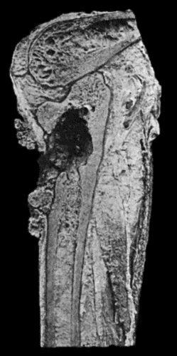
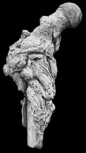
Fig. 153.—Epitheliomatous Ulcer of Leg with direct extension to Tibia.
(Lord Lister's specimen. Anatomical Museum, University of Edinburgh.)
Secondary cancer is a comparatively common disease, and, as in metastases in other tissues, the secondary growths resemble the parent tumour. The soft forms grow rapidly, and eat away the bone, without altering its shape or form. In slowly growing forms there may be considerable formation of imperfectly formed bone, often deficient in lime salts; this condition may be widely diffused throughout the skeleton, and, as it is associated with softening and bending of the bones, it is known as cancerous osteomalacia. Secondary cancer of bone is attended with pain, or it suddenly attracts notice by the occurrence of pathological fracture—as, for example, in the shaft of the femur or humerus. In the vertebræ, it is attended with a painful form of paraplegia, which may involve the lower or all four extremities. On the other hand, the disease may show itself clinically as a tumour of bone, which may attain a considerable size, and may be mistaken for a sarcoma, unless the existence of the primary cancer is discovered.
The cancers most liable to give rise to metastasis in bone are those of the breast, liver, uterus, prostate, colon, and rectum; hyper-nephroma of the kidney may also give rise to metastases in bone.
Secondary tumours derived from the thyreoid gland require special mention, because they are peculiar in that neither the primary growth in the thyreoid nor the secondary growth in the bones is necessarily malignant. They are therefore amenable to operative treatment.
Secondary sarcoma, whether derived from a primary growth in the bone or in the soft parts, is much rarer than secondary cancer. Its removal by operation is usually contra-indicated, but we have known of cases terminating fatally in which the section revealed only one metastasis, the removal of which would have benefited the patient.
In all of these conditions, examination of the bones with the X-rays gives valuable information and often disclose unsuspected metastases.
Cancer of Bone resulting from Direct Extension from Soft Parts.—In this group there are also two clinical types. The first is met with in relation to epithelioma of a mucous surface—for example, the palate, tongue, gums, antrum, frontal sinus, auditory meatus, or middle ear. They will be described under these special regions.
The second type is met with in relation to epithelioma occurring in a sinus, the sequel of suppurative osteomyelitis, compound fracture, or tuberculous disease. The patient has usually had a discharging sinus for a great number of years: we have known it to last as many as fifty. The epithelioma originates at the skin orifice of the sinus, and spreads to the bone and into its interior, where the progress of the cancer is resisted by dense bone, which obliterates the medullary canal. Although its progress is slow, the infiltration of the bone is usually more extensive than appears externally. It is recognised clinically by the characteristic cauliflower growth at the orifice of the sinus, and by the offensive nature of the discharge. A similar epithelioma may arise in connection with a chronic ulcer of the leg. The cancer may infect the femoral lymph glands. The operative treatment is influenced by the extent of the disease in the soft parts overlying the bone, and consists in wide removal of the diseased tissues and resection of the bone, or in amputation.
Cysts of Bone.—With the exception of hydatid cysts, cysts in the interior of bone are the result of the liquefaction of solid tissue; this may be that of chondroma, myeloma, or sarcoma, but more commonly of the marrow in osteomyelitis fibrosa.