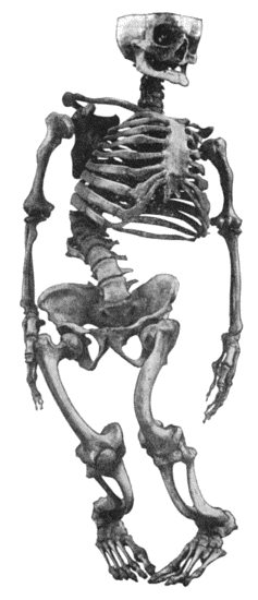
Fig. 133.—Skeleton of Rickety Dwarf, known as "Bowed Joseph," leader of the Meal Riots in Edinburgh, who died in 1780.
(Anatomical Museum, University of Edinburgh.)
Rickets or rachitis is a constitutional disease associated with disturbance of nutrition, and attended with changes in the skeleton. The disease is most common and most severe among the children of the poorer classes in large cities, who are improperly fed and are brought up in unhealthy surroundings. There is evidence that the most important factors in the causation of rickets are ill-health of the mother during pregnancy, and the administration to the child after its birth of food which is defective in animal fat, proteids, and salts of lime, or which contains these in such a form that they are not readily assimilated. The occurrence of the disease is favoured, and its features are aggravated, by imperfect oxygenation of the blood as the result of a deficiency of fresh air and sunlight, want of exercise, and by other conditions which prevail in the slums of large towns.
Pathological Anatomy.—The most striking feature is the softness (malacia) of the bones, due to excessive absorption of osseous tissue, and the formation of an imperfectly calcified tissue at the sites of ossification. The affected bones lose their rigidity, so that they are bent under the weight of the body, by the traction of muscles, and by other mechanical forces.
The periosteum is thick and vascular, and when detached carries with it plates and spicules of soft porous bone. The new bone may be so abundant that it forms a thick crust on the surface, and in the flat bones of the skull this may be heaped up in the form of bosses or ridges resembling those ascribed to inherited syphilis.
In the epiphysial cartilages and at the ossifying junctions, all the processes concerned in ossification, excepting the deposition of lime salts, occur to an exaggerated degree. The cartilage of the epiphysial disc proliferates actively and irregularly, so that it becomes softer, thicker, and wider, and gives rise to a visible swelling, best seen at the lower end of the radius and lower end of the tibia, and at the costo-chondral junctions where the series of beaded swellings is known as the “rickety rosary.”
The ossifying zone is increased in depth; the marrow is abnormally vascular; and the new bone that is formed is imperfectly calcified. The result is that the bones may never attain their normal length, and they remain stunted throughout life as in rickety dwarfs (Fig. 133), or the shafts may grow unequally and come to deviate from their normal axes as in knock-knee and bow-knee.
Fig. 133.—Skeleton of Rickety Dwarf, known as "Bowed Joseph," leader of the Meal Riots in Edinburgh, who died in 1780.
(Anatomical Museum, University of Edinburgh.)
These changes are well brought out in skiagrams; instead of the well-defined narrow line which represents the epiphysial cartilage, there is an ill-defined, blurred zone of considerable depth.
In the shafts of the long bones, owing to the excessive absorption of bone, the cortex becomes porous, the spongy bone is rarefied, and the bones readily bend or break under mechanical influences. When the disease is arrested, a process of repair sets in which often results in the bones becoming denser and heavier than normal. In the flat bones of the skull, the absorption may result in the entire disappearance of areas of bone, leaving a membrane which dimples like thin cardboard under the pressure of the finger—a condition known as craniotabes.
Changes in the Skeleton before the Child is able to walk.—The fontanelles remain open until the end of the second year or longer, and the frontal and parietal eminences are unduly prominent. There is sometimes hydrocephalus, and the head is characteristically enlarged. The jaws are altered so that while the upper jaw is contracted into the shape of a V, the lower jaw is square instead of rounded in outline, and the teeth do not oppose one another. In the thorax, the chief feature may be the beading at the costo-chondral junctions, principally of the fifth and sixth ribs or its walls may be contracted, particularly if respiration is interfered with as a result of bronchial catarrh or adenoids. The contraction may take the form of a vertical groove on each side, or of a horizontal groove at the level of the upper end of the xiphi-sternum; when the sternum and cartilages form a projection in front, the deformity is known as “pigeon-breast.”
The spine may be curved backwards—kyphosis—throughout its whole extent or only in one part; or it may be curved to one side—scoliosis.
In the limbs, the prominent features are the deficient growth in length of the long bones, the enlargements at the epiphysial junctions, and the bending, and occasional greenstick fracture, of the shafts. The degree of enlargement of the epiphysial junctions is directly proportionate to the amount of movement to which the bone is subjected (John Thomson). The curves at this stage depend on the attitude of the child while sitting or being carried—for example, the arm bones become bent in children who paddle about the floor with the aid of their arms; and in a child who lies on its back with the lower limbs everted, the weight of the limb may lead to curvature of the neck of the femur—coxa vara. The clavicle or humerus may sustain greenstick fracture from the child being lifted by the arms; the femur, by a fall. From the extreme laxity of the ligaments, the joints can be moved beyond the normal limits, and the child is often observed to twist its limbs into abnormal attitudes.
In Children who have walked.—In these children the most important deformities occur in the spine, pelvis, and lower extremities, and result for the most part from yielding of the softened bones under the weight of the body. Scoliosis is the usual type of spinal curvature, and in extreme cases it may lead to a pronounced form of hump-back. The pelvis may remain small (justo-minor pelvis), or it may be contracted in the sagittal plane (flat pelvis); when the bones are unusually soft, the acetabular portions are pushed inwards by the femora bearing the weight of the body, and the pelvis assumes the shape of a trefoil, as in the malacia of women. The shaft of the femur is curved forwards and laterally; the bones of the leg laterally as in bow-leg, or forwards, or forwards and laterally just above the ankle. The deformities at the knee (genu valgum, genu varum, and genu recurvatum), and at the hip (coxa vara), will be described in the volume dealing with the Extremities.
The majority of cases seen in surgical practice suffer from the deformities resulting from rickets rather than from the active disease. The examination of a large series of children at different ages shows that the deformities become less and less frequent with each year. Those who recover may ultimately show no trace of rickets, and this is especially true of children who grow at the average rate; in those, however, in whom growth is retarded, especially from the fifth to the seventh year, the deformities are apt to be permanent. It may be noted that the scoliosis due to rickets has little tendency towards recovery.
Treatment.—The treatment of the disease consists in regulating the diet, improving the surroundings, and preventing deformity. Phosphorus in doses of 100th grain may be given dissolved in cod-liver oil, and preparations of iron and lime may be added with advantage. To avoid those postures which predispose to deformities, the child should lie as much as possible. In the well-to-do classes this is readily accomplished by the aid of a nurse and the use of a perambulator. In hospital out-patients the child is kept off its feet by the use of a light wooden splint applied to the lateral aspect of each lower extremity, and extending from the pelvis to 6 inches beyond the sole.
When deformities are already present, the treatment depends upon whether or not there is any prospect of the bone straightening naturally. Under five years of age this may, as a rule, be confidently expected; the child should be kept off its feet, and the limbs bathed and massaged. In children of five or six and upwards, the prospect of natural straightening is a diminishing one, and it is more satisfactory to correct the deformity by operation. In rickety curvature of the spine, the child should lie on a firm mattress, or, to allow of its being taken into the open air, upon a double Thomas' splint extending from the occiput to the heels; the muscles acting on the trunk should be braced up by massage and appropriate exercises.
Late Rickets or Rachitis Adolescentium is met with at any age from nine to seventeen, and is generally believed to be due to a recrudescence of rickets which had been present in childhood. The disease is not attended with any disturbance of the general health; the pathological changes are the same as in infantile rickets, but are for the most part confined to the ossifying junctions, especially those which are most active during adolescence, for example at the knee-joint. The patient is easily tired, complains of pain in the bones, and, unless care is taken, deformity is liable to ensue. There can be no doubt that adolescent rickets plays an important part in the production of the deformities which occur at or near puberty, especially knock-knee and bow-knee.
Scurvy-Rickets or Infantile Scurvy.—This disease, described by Barlow and Cheadle, is met with in infants under two years who have been brought up upon sterilised or condensed milk and other proprietary foods, and is most common in the well-to-do classes. The hæmorrhages, which are so characteristic of the disease, are usually preceded for some weeks by a cachectic condition, with listlessness and debility and disinclination for movement. Very commonly the child ceases to move one of his lower limbs—pseudo-paralysis—and screams if it is touched; a swelling is found over one of the bones, usually the femur, accompanied by exquisite tenderness; the skin is tense and shiny, and there may be some œdema. These symptoms are due to a sub-periosteal hæmorrhage, and associated with this there may be crepitus from separation of an epiphysis, rarely from fracture of the shaft of the bone. X-ray photographs show enlargement of the bone, the periosteum being raised from the shaft and new bone formed in relation to it. Hæmorrhages also occur into the skin, presenting the appearance of bruises, into the orbit and conjunctiva, and from the mucous membranes.
The treatment consists in correcting the errors in diet. The infant should have a wet nurse or a plentiful supply of cow's milk in its natural state. Anti-scorbutics in the form of orange, lemon, or grape juice, and of potatoes bruised down in milk, may be given.
Osteomalacia.—The term osteomalacia includes a group of conditions, closely allied to rickets, in which the bones of adults become soft and yielding, so that they are unduly liable to bend or break.
One form occurs in pregnant and puerperal women, affecting most commonly the pelvis and lumbar vertebræ, but sometimes the entire skeleton. The lime salts are absorbed, the bones lose their rigidity and bend under the weight of the body and other mechanical influences, with the result that gross deformities are produced, particularly in the pelvis, the lumbar spine, and the hip-joints.
Neuropathic forms occur in certain chronic diseases of the brain and cord; in some cases the bones lose their lime salts and bend, in others they become brittle.
Osteomalacia associated with New Growths in the Skeleton.—When secondary cancer is widely distributed throughout the skeleton, it is associated with softening of the bones, as a result of which they readily bend or break, and after death are easily cut with a knife. In the disease known as multiple myeloma, the interior of the ribs, sternum, and bodies of the vertebræ is occupied by a reddish gelatinous pulp, the structure of which resembles sarcoma; the bones are reduced to a mere shell, and may break on the slightest pressure; the urine contains albumose, a substance resembling albumen but coagulating at a comparatively low temperature (140° F.), and the coagulum is re-dissolved on boiling, and it is readily precipitated by hydrochloric acid (Bence-Jones).
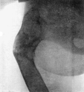
Ostitis Deformans—Paget's Disease of Bone.—This rare disease was first described by Sir James Paget in 1877. In the early stages, the marrow is transformed into a vascular connective tissue; its bone-eating functions are exaggerated, and the framework of the bone becomes rarefied, so that it bends under pressure as in osteomalacia. In course of time, however, new bone is formed in great abundance; it is at first devoid of lime salts, but later becomes calcified, so that the bones regain their rigidity. This formation of new bone is much in excess of the normal, the bones become large and bulky, their surfaces rough and uneven, their texture sclerosed in parts, and the medullary canal is frequently obliterated. These changes are well brought out in X-ray photographs. The curving of the long bones, which is such a striking feature of the disease, may be associated with actual lengthening, and the changes are sometimes remarkably symmetrical (Fig. 135). The bones forming the cranium may be enormously thickened, the sutures are obliterated, the distinction into tables and diploë is lost, and, while the general texture is finely porous, there may be areas as dense as ivory (Fig. 134).
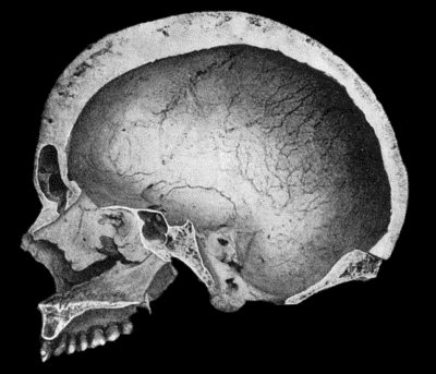
Fig. 134.—Changes in the Skull resulting from Ostitis Deformans.
(Anatomical Museum, University of Edinburgh.)
Clinical Features.—The disease is usually met with in persons over fifty years of age. It is insidious in its onset, and, the patient's attention may be first attracted by the occurrence of vague pains in the back or limbs; by the enlargement and bending of such bones as the tibia or femur; or by a gradual increase in the size of the head, necessitating the wearing of larger hats. When the condition is fully developed, the attitude and general appearance are eminently characteristic. The height is diminished, and, owing to the curving of the lower limbs and spine, the arms appear unnaturally long; the head and upper part of the spine are bent forwards; the legs are held apart, slightly flexed at the knees, and are rotated out as well as curved; the whole appearance suggests that of one of the large anthropoid apes. The muscles of the limbs may waste to such an extent as to leave the large, curved, misshapen bones covered only by the skin (Fig. 135). In the majority of cases the bones of the lower extremities are much earlier and more severely affected than those of the upper extremity, but the capacity of walking is usually maintained even in the presence of great deformity. In a case observed by Byrom Bramwell, the patient suffered from a succession of fractures over a period of years.
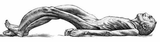
Fig. 135.—Cadaver, illustrating the alterations in the Lower Limbs resulting from Ostitis Deformans.
The disease may last for an indefinite period, the general health remaining long unaffected. In a considerable number of the recorded cases one of the bones became the seat of sarcoma.
Osteomyelitis Fibrosa.—This comparatively rare disease, which was first described by Recklinghausen, presents many interesting features. Because of its causing deformities of the bones and an undue liability to fracture, and being chiefly met with in adolescents, it is regarded by some authors as a juvenile form of Paget's disease. It may be diffused throughout the skeleton—we have seen it in the skull and in the bones of the extremities—or it may be confined to a single bone, usually the femur, or, what is more remarkable, the condition may affect a portion only of the shaft of a long bone and be sharply defined from the normal bone in contact with it.
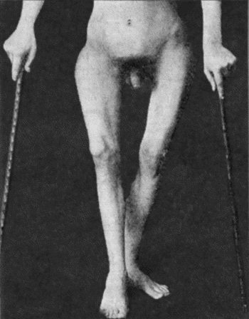
Fig. 136.—Osteomyelitis Fibrosa affecting Femora in a man æt. 19. The curving of the bones is due to multiple fractures.
On longitudinal section of a long bone during the active stage of the disease, the marrow is seen to be replaced by a vascular young connective tissue which encroaches on the surrounding spongy bone, reducing it to the slenderest proportions; the formation of bone from the periosteum does not keep pace with the absorption and replacement going on in the interior, and the cortex may be reduced to a thin shell of imperfectly calcified bone which can be cut with a knife. The young connective tissue which replaces the marrow is not unlike that seen in osteomalacia; it is highly vascular and may show hæmorrhages of various date; there are abundant giant cells of the myeloma type, and degeneration and liquefaction of tissue may result in the formation of cysts, which, when they constitute a prominent feature, are responsible for the name—osteomyelitis fibrosa cystica—sometimes applied to the condition.
It would appear that most of the recorded cases of cysts of bone owe their origin to this disease, while the abundance of giant cells with occasional islands of cartilage in the wall of such cysts is responsible for the view formerly held that they owed their origin to the liquefaction of a solid tumour, such as a myeloma, a chondroma, or even a sarcoma. Although the tissue elements in this disease resemble those of a new growth arising in the marrow, they differ in their arrangement and in their method of growth; there is no tendency to erupt through the cortex of the bone, to invade the soft parts, or to give rise to secondary growths.
Clinical Features.—The onset of the disease is insidious, and attention is usually first directed to it by the occurrence of fracture of the shaft of one of the long bones—usually the femur—from violence that would be insufficient to break a healthy bone. Apart from fracture, the great increase in the size of one of the long bones and its uneven contour are sufficiently remarkable to suggest examination with the X-rays, by means of which the condition is at once recognised. A systematic examination of the other long bones will often reveal the presence of the disease at a stage before the bone is altered externally.
Symmetrical bossing of the skull was present in the case shown in Figs. 136 and 137, and there were also scattered patches of brown pigmentation of the skin of the face, neck, and trunk, similar to those met with in generalised neuro-fibromatosis. Apart from fracture, the disease is recognised by the thickening and usually also by the curving of the shafts of the long bones. It is easy to understand the curvature of bones that have passed through a soft stage and also of those that have been broken and badly united, but it is difficult to account for the curvatures that have no such cause; for example, we have seen marked curve of the radius in a forearm of which the ulna was quite straight. The curvature probably resulted from exaggerated growth in length.
The X-ray appearances vary with the stage of the malady, not estimated in time, for the condition is chronic and may become stationary, but according to whether it is progressive or undergoing repair. The shadow of the bone presents a poor contrast to the soft parts, and no trace of its original architecture; in extreme cases the shadow of the femur resembles an unevenly filled sausage (Fig. 137); there is no cortical layer, the interior shows no trabecular structure, and some of the many clear areas are probably cysts. The condition extends right up to the articular cartilage, or, in the case of adolescent bones, up to the epiphysial cartilage.
Prognosis.—The condition does not appear to affect the general health. The future is concerned with the local conditions, and, especially in the case of the femur, with its liability to fracture; so far as we know there is no time limit to this.
Treatment is confined to protecting the affected bone—usually the femur—from injury. Operative treatment may be required for lameness due to a badly united fracture.
Neuropathic Atrophy of Bone.—The conditions included under this heading occur in association with diseases of the nervous system.
Most importance attaches to the fragility of the bones met with in general paralysis of the insane, locomotor ataxia, and other chronic diseases of the brain and spinal cord. The bones are liable to be fractured by forces which would be insufficient to break a healthy bone. In locomotor ataxia the fractures affect especially the bones of the lower extremity, and may occur before there are any definite nerve symptoms, but they are more often met with in the ataxic stage, when the abrupt and uncontrolled movements of the limbs may play a part in their causation. They may be unattended with pain, and may fail to unite; when repair does take place, it is sometimes attended with an excessive formation of callus. Joint lesions of the nature of Charcot's disease may occur simultaneously with the alterations in the bones. In syringomyelia pathological fracture is not so frequent as in locomotor ataxia; it is more likely to occur in the bones of the upper extremity, and especially in the humerus. In some cases of epilepsy the bones break when the patient falls in a fit, and there is usually an exaggerated amount of comminution.
In these affections the bones present no histological or chemical alterations, and the X-ray shadow does not differ from the normal. It is maintained, therefore, that the disposition to fracture does not depend upon a fragility of the bone, but on the loss of the muscular sense and of common sensation in the bones, as a result of which there is an inability properly to throw the muscles into action and dispose the limbs so as to place them under the most favourable conditions to meet external violence.
Osteogenesis Imperfecta, Fragilitas Ossium, or Congenital Osteopsathyrosis.—These terms are used to describe a condition in which an undue fragility of the bones dates from intra-uterine life. It may occur in several members of the same family. In severe cases, intra-uterine fractures occur, and during parturition fresh fractures are almost sure to be produced, so that at birth there is a combination of recent fractures and old fractures united and partly united, with bendings and thickenings of the bones. Large areas of the cranial vault may remain membranous.
After birth the predisposition to fracture continues, the bones are easily broken, the fractures are attended with little or no pain, the crepitus is soft, and although union may take place, it may be delayed and be attended with excess of callus. Cases have been observed in which a child has sustained over a hundred fractures.
The bones show a feeble shadow with the X-rays, and appear thin and atrophied; the medullary canal is increased at the expense of the cortex.
In young infants in whom multiple fractures occur the prognosis as to life is unfavourable, and no satisfactory treatment of the disease has been formulated. If the patient survives, the tendency to fracture gradually disappears.
Hypertrophic Pulmonary Osteo-Arthropathy.—This condition, which was described by Marie in 1890, is secondary to disease in the chest, such as chronic phthisis, empyema, bronchiectasis, or sarcoma of the lung. There is symmetrical enlargement and deformity of the hands and feet; the shafts of the bones are thickened, and the soft tissues of the terminal segments of the digits hypertrophied. The fingers come to resemble drum-sticks, and the thumb the clapper of a bell. The nails are convex, and incurved at their free ends, suggesting a resemblance to the beak of a parrot. There is also enlargement of the lower ends of the bones of the forearm and leg, and effusion into the wrist and ankle-joints. Skiagrams of the hands and feet show a deposit of new bone along the shafts of the phalanges.