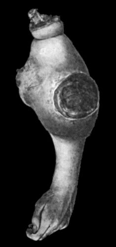
Fig. 55.—Recurrent Sarcoma of Sciatic Nerve in a woman æt. 27. Recurrence twenty months after removal of primary growth.
The term sarcoma is applied to any connective-tissue tumour which exhibits malignant characters. The essential structural feature is the predominance of the cellular elements over the intercellular substance or stroma, in which respect a sarcoma resembles the connective tissue of the embryo. The typical sarcoma consists chiefly of immature or embryonic connective tissue. It most frequently originates from fascia, intermuscular connective tissue, periosteum, bone-marrow, and skin, and forms a rounded or nodulated tumour which appears to be encapsulated, but the capsule merely consists of the condensed surrounding tissues, and usually contains sarcomatous elements. The consistence of the tumour depends on the nature and amount of the stroma, and on the presence of degenerative changes. The softer medullary forms are composed almost exclusively of cells; while the harder forms—such as the fibro-, chondro-, and osteo-sarcoma—are provided with an abundant stroma and are relatively poor in cells. Degenerative changes may produce areas of softening or liquefaction which result in the formation of cystic cavities in the interior of the tumour. The colour depends on the amount of blood in the tumour, and on the presence of the products of degeneration.
The blood vessels are usually represented by mere chinks or spaces between the cells. This peculiarity accounts for the facility with which hæmorrhage takes place into the substance of the tumour, the persistence of the bleeding when it is incised or ulcerates through the skin, and the readiness with which the sarcomatous cells are carried off and infect distant parts through the blood-stream. Sarcomas are devoid of lymphatics, and unless originating in lymphatic structures—for example, in the tonsil—they rarely infect the lymph glands. Minute portions of the tumour grow into the small veins, and, becoming detached, are transported by the blood-current to distant organs, where they are arrested in the capillaries and give rise to secondary growths. These are most frequently situated in the lungs, except when the primary growth lies within the territory of the portal circulation, in which case they occur in the liver. The secondary growths closely resemble the parent tumour. Sarcoma may invade an adjacent vein on such a scale that if the invading portion becomes detached it may constitute a dangerous embolus. This may be observed in sarcoma of the kidney, the growth taking place along the renal vein until it projects into the vena cava.
Fig. 55.—Recurrent Sarcoma of Sciatic Nerve in a woman æt. 27. Recurrence twenty months after removal of primary growth.
In its growth, a sarcoma compresses and destroys neighbouring parts, surrounds vessels and nerves, and may lead to destruction of the skin, either by invading it, or more commonly by causing sloughing from pressure. Inflammatory and suppurative changes may take place as a result of pyogenic infection following upon sloughing of the overlying skin or upon an exploratory incision. Once the skin is broken the tumour fungates through the opening. Sarcomas vary in malignancy, especially as regards rapidity of growth and capacity for dissemination. Certain of them, such as the so-called “recurrent fibroid of Paget,” grow comparatively slowly, and are only malignant in the sense that they tend to recur locally after removal; others—especially the more cellular ones—grow with extreme rapidity, and are early disseminated throughout the body, resembling in these respects the most malignant forms of cancer. They are usually solitary in the first instance, although primary multiple growths are occasionally met with in the skin and in the bones.
Many varieties of sarcoma are recognised, according to its structural peculiarities. Thus, in virtue of the size and character of the cells, we have the small round-celled and the large round-celled sarcoma, the small and the large spindle-celled, the giant-celled and the mixed-celled sarcoma. The lympho-sarcoma presents a structure similar to that of lymph-follicular tissue, and the alveolar sarcoma an arrangement of cells in alveoli resembling that seen in cancers. When there is a considerable amount of intercellular fibrous tissue, the tumour is called a fibro-sarcoma.
The term lymphangio-sarcoma is applied when the cells of the tumour are derived from the endothelium of lymph spaces and vessels. The angio-sarcomas are those in which blood vessels form a prominent element in the structure of the tumour. They are sometimes derived from innocent angiomas, and they may be so vascular as to pulsate and on auscultation yield a blowing murmur like an aneurysm. The glio-sarcoma, myxo-sarcoma, chondro-sarcoma, and myo-sarcoma are mixed forms which usually develop in pre-existing innocent tumours. The osteo-sarcoma is characterised by the formation in the tumour of bone, the medullary spaces being occupied by sarcomatous cells in place of marrow. The osteoid sarcoma is characterised by the formation of a tissue resembling bone but deficient in lime salts, and the petrifying sarcoma by the formation of calcified areas in the stroma. These varieties, although met with chiefly in the bones, may occur in soft tissues such as muscle, and in such organs as the mamma. The pigmented varieties include the chloroma, which is of a light-green colour, and the melanotic sarcoma, which is brown or black. The psammoma is a sarcoma containing a material resembling sand; it is chiefly met with in the membranes of the brain. The chordoma is a rare form of tumour originating from the remains of the notochord in the region of the spheno-occipital synchondrosis or in the sacro-coccygeal region.
Diagnosis of Sarcoma.—A sarcoma is to be differentiated from an inflammatory swelling such as results from tubercle, actinomycosis, or syphilis, from an innocent tumour, and from a cancer. The points on which the diagnosis is founded are discussed with the different tissues and organs.
Treatment.—The removal of the tumour by operation is the most reliable method of treatment; in order to be successful it must be undertaken before dissemination has taken place, and a considerable area of healthy tissue beyond the apparent margin of the growth must be removed, and in tumours near the surface of the body, the overlying skin also.
In order to prevent recurrence, a tube of radium, to which a silk thread is attached, is inserted into the space from which the tumour was removed; the thread is brought out at the drain-opening, and at the end of a week or ten days the tube of radium is removed by pulling on the thread. Radium causes a reaction in the tissues attended with exudation from the vessels, for the escape of which provision must be made. If radium is not available, the affected area is repeatedly exposed to the action of the X-rays as soon as the wound has healed. The employment of these measures has diminished to a remarkable degree the recurrence of sarcoma after operation.
It will readily be understood that the less thoroughly or radically the growth has been removed, the more do we depend upon radium or the X-rays for bringing about a permanent cure, and that in advanced cases of sarcoma and in cases in which, on account of their anatomical situation, removal by operation is necessarily incomplete, the prospect of cure is still more dependent on the use of radium or of the X-rays. Finally, there are cases in which removal by operation is impossible, the so-called inoperable sarcoma; a tube of radium, to which a silk thread is attached, is inserted into the substance of the tumour, either through an opening made by a large trocar, or, when necessary, by open dissection. A second tube of radium is placed upon the skin over the tumour and is secured there by a stitch or by a strip of plaster, thus securing a cross-fire action of the radium rays, both from within and without, as this is found to be much more efficacious in destroying or inhibiting the cellular elements of the growth. The tubes of radium are left in situ for from eight to fourteen days, according to the power of the radium employed, but are moved about every second day or so in order that every part of the tumour may be efficiently radiated. If the tumour shrinks in size after the use of radium and becomes operable, it should be removed before time is given it to resume its growth. It will depend upon the subsequent course of the disease, whether or not a second, or it may be even a third, application of radium will be required.
Where neither radium nor X-rays is available or applicable, recourse may be had to the injection of Coley's fluid, a preparation containing the mixed toxins of the streptococcus of erysipelas and the bacillus prodigiosus; or of selenium.