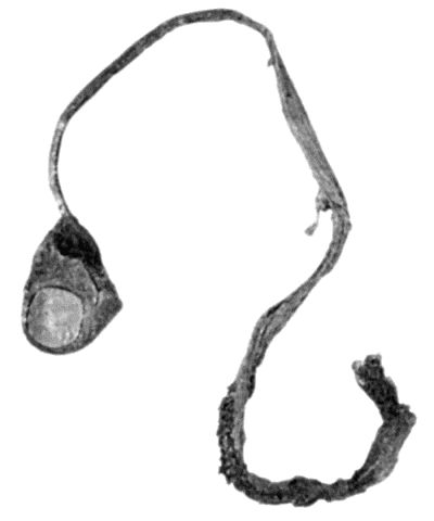
Fig. 108.—Avulsion of Tendon with Terminal Phalanx of Thumb.
(Surgical Museum, University of Edinburgh.)
Contusion of Muscle.—Contusion of muscle, which consists in bruising of its fibres and blood vessels, may be due to violence acting from without, as in a blow, a kick, or a fall; or from within, as by the displacement of bone in a fracture or dislocation.
The symptoms are those common to all contusions, and the patient complains of severe pain on attempting to use the muscle, and maintains an attitude which relaxes it. If the sheath of the muscle also is torn, there is subcutaneous ecchymosis, and the accumulation of blood may result in the formation of a hæmatoma.
Restoration of function is usually complete; but when the nerve supplying the muscle is bruised at the same time, as may occur in the deltoid, wasting and loss of function may be persistent. In exceptional cases the process of repair may be attended with the formation of bone in the substance of the muscle, and this may likewise impair its function.
A contused muscle should be placed at rest and supported by cotton wool and a bandage; after an interval, massage and appropriate exercises are employed.
Sprain and Partial Rupture of Muscle.—This lesion consists in overstretching and partial rupture of the fibres of a muscle or its aponeurosis. It is of common occurrence in athletes and in those who follow laborious occupations. It may follow upon a single or repeated effort—especially in those who are out of training. Familiar examples of muscular sprain are the “labourer's” or “golfer's back,” affecting the latissimus dorsi or the sacrospinalis (erector spinæ); the “tennis-player's elbow,” and the “sculler's sprain,” affecting the muscles and ligaments about the elbow; the “angler's elbow,” affecting the common origin of the extensors and supinators; the “sprinter's sprain,” affecting the flexors of the hip; and the “jumper's and dancer's sprain,” affecting the muscles of the calf. The patient complains of pain, often sudden in onset, of tenderness on pressure, and of inability to carry out the particular movement by which the sprain was produced. The disability varies in different cases, and it may incapacitate the patient from following his occupation or sport for weeks or, if imperfectly treated, even for months.
The treatment consists in resting the muscle from the particular effort concerned in the production of the sprain, in gently exercising it in other directions, in the use of massage, and the induction of hyperæmia by means of heat. In neglected cases, that is, where the muscle has not been exercised, the patient shrinks from using it and the disablement threatens to be permanent; it is sometimes said that adhesions have formed and that these interfere with the recovery of function. The condition may be overcome by graduated movements or by a sudden forcible movement under an anæsthetic. These cases afford a fruitful field for the bone-setter.
Rupture of Muscle or Tendon.—A muscle or a tendon may be ruptured in its continuity or torn from its attachment to bone. The site of rupture in individual muscles is remarkably constant, and is usually at the junction of the muscular and tendinous portions. When rupture takes place through the belly of a muscle, the ends retract, the amount of retraction depending on the length of the muscle, and the extent of its attachment to adjacent aponeurosis or bone. The biceps in the arm, and the sartorius in the thigh, furnish examples of muscles in which the separation between the ends may be considerable.
The gap in the muscle becomes filled with blood, and this in time is replaced by connective tissue, which forms a bond of union between the ends. When the space is considerable the connecting medium consists of fibrous tissue, but when the ends are in contact it contains a number of newly formed muscle fibres. In the process of repair, one or both ends of the muscle or tendon may become fixed by adhesions to adjacent structures, and if the distal portion of a muscle is deprived of its nerve supply it may undergo degeneration and so have its function impaired.
Rupture of a muscle or tendon is usually the result of a sudden, and often involuntary, movement. As examples may be cited the rupture of the quadriceps extensor in attempting to regain the balance when falling backwards; of the gastrocnemius, plantaris, or tendo-calcaneus in jumping or dancing; of the adductors of the thigh in gripping a horse when it swerves—“rider's sprain”; of the abdominal muscles in vomiting, and of the biceps in sudden movements of the arm. Sometimes the effort is one that would scarcely be thought likely to rupture a muscle, as in the case recorded by Pagenstecher, where a professional athlete, while sitting at table, ruptured his biceps in a sudden effort to catch a falling glass. It would appear that the rupture is brought about not so much by the contraction of the muscle concerned, as by the contraction of the antagonistic muscles taking place before that of the muscle which undergoes rupture is completed. The violent muscular contractions of epilepsy, tetanus, or delirium rarely cause rupture.
The clinical features are usually characteristic. The patient experiences a sudden pain, with the sensation of being struck with a whip, and of something giving way; sometimes a distant snap is heard. The limb becomes powerless. At the seat of rupture there is tenderness and swelling, and there may be ecchymosis. As the swelling subsides, a gap may be felt between the retracted ends, and this becomes wider when the muscle is thrown into contraction. If untreated, a hard, fibrous cord remains at the seat of rupture.
Treatment.—The ends are approximated by placing the limb in an attitude which relaxes the muscle, and the position is maintained by bandages, splints, or special apparatus. When it is impossible thus to approximate the ends satisfactorily, the muscle or tendon is exposed by incision, and the ends brought into accurate contact by catgut sutures. This operation of primary suture yields the most satisfactory results, and is most successful when it is done within five or six days of the accident. Secondary suture after an interval of months is rendered difficult by the retraction of the ends and by their adhesion to adjacent structures.
Rupture of the biceps of the arm may involve the long or the short head, or the belly of the muscle. Most interest attaches to rupture of the long tendon of origin. There is pain and tenderness in front of the upper end of the humerus, the patient is unable to abduct or to elevate the arm, and he may be unable to flex the elbow when the forearm is supinated. The long axis of the muscle, instead of being parallel with the humerus, inclines downwards and outwards. When the patient is asked to contract the muscle, its belly is seen to be drawn towards the elbow.
The adductor longus may be ruptured, or torn from the pubes, by a violent effort to adduct the limb. A swelling forms in the upper and medial part of the thigh, which becomes smaller and harder when the muscle is thrown into contraction.
The quadriceps femoris is usually ruptured close to its insertion into the patella, in the attempt to avoid falling backwards. The injury is sometimes bilateral. The injured limb is rendered useless for progression, as it suddenly gives way whenever the knee is flexed. Treatment is conducted on the same lines as in transverse fracture of the patella; in the majority of cases the continuity of the quadriceps should be re-established by suture within five or six days of the accident.
The tendo calcaneus (Achillis) is comparatively easily ruptured, and the symptoms are sometimes so slight that the nature of the injury may be overlooked. The limb should be put up with the knee flexed and the toes pointed. This may be effected by attaching one end of an elastic band to the heel of a slipper, and securing the other to the lower third of the thigh. If this is not sufficient to bring the ends into apposition they should be approximated by an open operation.
The plantaris is not infrequently ruptured from trivial causes, such as a sudden movement in boxing, tennis, or hockey. A sharp stinging pain like the stroke of a whip is felt in the calf; there is marked tenderness at the seat of rupture, and the patient is unable to raise the heel without pain. The injury is of little importance, and if the patient does not raise the heel from the ground in walking, it is recovered from in a couple of weeks or so, without it being necessary to lay him up.
Hernia of Muscle.—This is a rare condition, in which, owing to the fascia covering a muscle becoming stretched or torn, the muscular substance is protruded through the rent. It has been observed chiefly in the adductor longus. An oval swelling forms in the upper part of the thigh, is soft and prominent when the muscle is relaxed, less prominent when it is passively extended, and disappears when the muscle is thrown into contraction. It is liable to be mistaken, according to its situation, for a tumour, a cyst, a pouched vein, or a femoral or obturator hernia. Treatment is only called for when it is causing inconvenience, the muscle being exposed by a suitable incision, the herniated portion excised, and the rent in the sheath closed by sutures.
Dislocation of Tendons.—Tendons which run in grooves may be displaced as a result of rupture of the confining sheath. This injury is met with chiefly in the tendons at the ankle and in the long tendon of the biceps.
Dislocation of the peronei tendons may occur, for example, from a violent twist of the foot. There is severe pain and considerable swelling on the lateral aspect of the ankle; the peroneus longus by itself, or together with the brevis, can be felt on the lateral aspect or in front of the lateral malleolus; the patient is unable to move the foot. By a little manipulation the tendons are replaced in their grooves, and are retained there by a series of strips of plaster. At the end of three weeks massage and exercises are employed.
In other cases there is no history of injury, but whenever the foot is everted the tendon of the peroneus longus is liable to be jerked forwards out of its groove, sometimes with an audible snap. The patient suffers pain and is disabled until the tendon is replaced. Reduction is easy, but as the displacement tends to recur, an operation is required to fix the tendon in its place. An incision is made over the tendon; if the sheath is slack or torn, it is tightened up or closed with catgut sutures; or an artificial sheath is made by raising up a quadrilateral flap of periosteum from the lateral aspect of the fibula, and stitching it over the tendon.
Similarly the tibialis posterior may be displaced over the medial malleolus as a result of inversion of the foot.
The long tendon of the biceps may be dislocated laterally—or more frequently medially—as a result of violent or repeated rotation movements of the arm, such as are performed in wringing clothes. The patient is aware of the displacement taking place, and is unable to extend the forearm until the displaced tendon has been reduced by abducting the arm. In recurrent cases the patient may be able to dislocate the tendon at will, but the disability is so inconsiderable that there is rarely any occasion for interference.
Wounds of Muscles and Tendons.—When a muscle is cut across in a wound, its ends should be brought together with sutures. If the ends are allowed to retract, and especially if the wound suppurates, they become united by scar tissue and fixed to bone or other adjacent structure. In a limb this interferes with the functions of the muscle; in the abdominal wall the scar tissue may stretch, and so favour the development of a ventral hernia.
Tendons may be cut across accidentally, especially in those wounds so commonly met with above the wrist as a result, for example, of the hand being thrust through a pane of glass. It is essential that the ends should be sutured to each other, and as the proximal end is retracted the original wound may require to be enlarged in an upward direction. When primary suture has been omitted, or has failed in consequence of suppuration, the separated ends of the tendon become adherent to adjacent structures, and the function of the associated muscle is impaired or lost. Under these conditions the operation of secondary suture is indicated.
A free incision is necessary to discover and isolate the ends of the tendon; if the interval is too wide to admit of their being approximated by sutures, means must be taken to lengthen the tendon, or one from some other part may be inserted in the gap. A new sheath may be provided for the tendon by resecting a portion of the great saphenous vein.
Injuries of the tendons of the fingers are comparatively common. One of the best known is the partial or complete rupture of the aponeurosis of the extensor tendon close to its insertion into the terminal phalanx—drop- or mallet-finger. This may result from comparatively slight violence, such as striking the tip of the extended finger against an object, or the violence may be more severe, as in attempting to catch a cricket ball or in falling. The terminal phalanx is flexed towards the palm and the patient is unable to extend it. The treatment consists in putting up the finger with the middle joint strongly flexed. In neglected cases, a perfect functional result can only be obtained by operation; under a local anæsthetic, the ruptured tendon is exposed and is sutured to the base of the phalanx, which may be drilled for the passage of the sutures.
Subcutaneous rupture of one or other of the digital tendons in the hand or at the wrist can be remedied only by operation. When some time has elapsed since the accident, the proximal end may be so retracted that it cannot be brought down into contact with the distal end, in which case a slip may be taken from an adjacent tendon; in the case of one of the extensors of the thumb, the extensor carpi radialis longus may be detached from its insertion and stitched to the distal end of the tendon of the thumb.
Subcutaneous rupture of the tendon of the extensor pollicis longus at the wrist takes place just after its emergence from beneath the annular ligament; the actual rupture may occur painlessly, more frequently a sharp pain is felt over the back of the wrist. The prominence of the tendon, which normally forms the ulnar border of the snuff-box, disappears. This lesion is chiefly met with in drummer-boys and is the cause of drummer's palsy. The only chance of restoring function is in uniting the ruptured tendon by open operation.

Fig. 108.—Avulsion of Tendon with Terminal Phalanx of Thumb.
(Surgical Museum, University of Edinburgh.)
Avulsion of Tendons.—This is a rare injury, in which the tendons of a finger or toe are torn from their attachments along with a portion of the digit concerned. In the hand, it is usually brought about by the fingers being caught in the reins of a runaway horse, or being seized in a horse's teeth, or in machinery. It is usually the terminal phalanx that is separated, and with it the tendon of the deep flexor, which ruptures at its junction with the belly of the muscle (Fig. 108). The treatment consists in disinfecting the wound, closing the tendon-sheath, and trimming the mutilated finger so as to provide a useful stump.