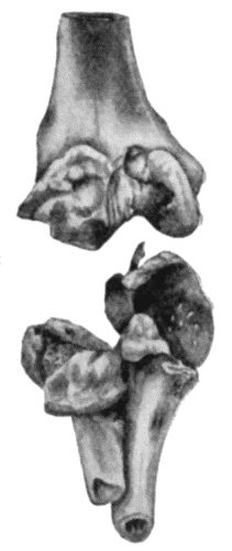
Fig. 157.—Arthritis Deformans of Elbow, showing destruction of articular surfaces and masses of new bone around the articular margins.
(Anatomical Museum, University of Edinburgh.)
Gout.—Arthritis Urica.—One of the manifestations of gout is that certain joints are liable to attacks of inflammation associated with the deposit of a chalk-like material composed of sodium biurate, chiefly in the matrix of the articular cartilage, it may be in streaks or patches towards the central area of the joint, or throughout the entire extent of the cartilage, which appears as if it had been painted over with plaster of Paris. As a result of this uratic infiltration, the cartilage loses its vitality and crumbles away, leading to the formation of what are known as gouty ulcers, and these may extend through the cartilage and invade the bone. The deposit of urates in the synovial membrane is attended with effusion into the joint and the formation of adhesions, while in the ligaments and peri-articular structures it leads to the formation of scar tissue. The metatarso-phalangeal joint of the great toe, on one or on both sides, is that most frequently affected. The disease is met with in men after middle life, and while common enough in England and Ireland, is almost unknown in hospital practice in Scotland.
The clinical features are characteristic. There is a sudden onset of excruciating pain, usually during the early hours of the morning, the joint becomes swollen, red, and glistening, with engorgement of the veins and some fever and disturbance of health and temper. In the course of a week or ten days there is a gradual return to the normal. Such attacks may recur only once a year or they may be more frequent; the successive attacks tend to become less acute but last longer, and the local phenomena persist, the joint remaining permanently swollen and stiff. Masses of chalk form in and around the joint, and those in the subcutaneous tissue may break through the skin, forming indolent ulcers with exposure of the chalky masses (tophi). The hands may become seriously crippled, especially when the tendon sheaths and bursæ also are affected; the crippling resembles that resulting from arthritis deformans but it differs in not being symmetrical.
The local treatment consists in employing soothing applications and a Bier's bandage for two or three hours twice daily while the symptoms are acute; later, hot-air baths, massage, and exercises are indicated. It is remarkable how completely even the most deformed joints may recover their function. Dietetic and medicinal treatment must also be employed.
Chronic Rheumatism.—This term is applied to a condition which sometimes follows upon acute articular rheumatism in persons presenting a family tendency to acute rheumatism or to inflammations of serous membranes, and manifesting other evidence of the rheumatic taint, such as chorea or rheumatic nodules.
The changes in the joints involve almost exclusively the synovial membrane and the ligaments; they consist in cellular infiltration and exudation, resulting in the formation of new connective tissue which encroaches on the cavity of the joint and gives rise to adhesions, and by contracting causes stiffness and deformity. The articular cartilages may subsequently be transformed into connective tissue, with consequent fibrous ankylosis and obliteration of the joint. The bones are affected only in so far as they undergo fatty atrophy from disuse of the limb, or alteration in their configuration as a result of partial dislocation. Osseous ankylosis may occur, especially in the small joints of the hand and foot.
The disease is generally poly-articular and may be met with in childhood and youth as well as in adult life. In some cases pain is so severe that the patient resists the least attempt at movement. In others, the joints, although stiff, can be moved but exhibit pronounced crackings. When there is much connective tissue formed in relation to the synovial membrane, the joint is swollen, and as the muscles waste above and below, the swelling is spindle-shaped. Subacute exacerbations occur from time to time, with fever and aggravation of the local symptoms and implication of other joints. After repeated recurrences, there is ankylosis with deformity, the patient becoming a helpless cripple. On account of the tendency to visceral complications, the tenure of life is uncertain.
From the nature of the disease, treatment is for the most part palliative. Salicylates are only of service during the exacerbations attended with pyrexia. The application of soda fomentations, turpentine cloths, or electric or hot-air baths may be useful. Improvement may result from the general and local therapeutics available at such places as Bath, Buxton, Harrogate, Strathpeffer, Wiesbaden, or Aix. In selected cases, a certain measure of success has followed operative interference, which consists in a modified excision. The deformities resulting from chronic rheumatism are but little amenable to surgical treatment, and forcible attempts to remedy stiffness or deformity are to be avoided.
Arthritis Deformans (Osteo-arthritis, Rheumatoid Arthritis, Rheumatic Gout, Malum Senile, Traumatic or Mechanical Arthritis).—Under the term arthritis deformans, which was first employed by Virchow, it is convenient to include a number of joint affections which have many anatomical and clinical features in common.
Fig. 157.—Arthritis Deformans of Elbow, showing destruction of articular surfaces and masses of new bone around the articular margins.
(Anatomical Museum, University of Edinburgh.)
The disease is widely distributed in the animal kingdom, both in domestic species and in wild animals in the natural state such as the larger carnivora and the gorilla; evidence of it has also been found in the bones of animals buried with prehistoric man.
The morbid changes in the joints present a remarkable combination of atrophy and degeneration on the one hand and overgrowth on the other, indicating a profound disturbance of nutrition in the joint structures. The nature of this disturbance and its etiology are imperfectly known. By many writers it is believed to depend upon some form of auto-intoxication, the toxins being absorbed from the gastro-intestinal tract, and those who suffer are supposed to possess what has been called an “arthritic diathesis.”
The localisation of the disease in a particular joint may be determined by several factors, of which trauma appears to be the most important. The condition is frequently observed to follow, either directly or after an interval, upon a lesion which involves gross injury of the joint or of one of the neighbouring bones. It occurs with greater frequency after repeated minor injuries affecting the joint and its vicinity, such as sprains and contusions, and particularly those sustained in laborious occupations. This connection between trauma and arthritis deformans led Arbuthnot Lane to apply to it the term traumatic or trade arthritis.
The traumatic or strain factor in the production of the disease may be manifested in a less obvious fashion. In the lower extremity, for example, any condition which disturbs the static equilibrium of the limb as a whole would appear to predispose to the disease in one or other of the joints. The static equilibrium may be disturbed by such deformities as flat-foot or knock-knee, and badly united fractures of the lower extremity. In hallux valgus, the metatarso-phalangeal joint of the great toe undergoes changes characteristic of arthritis deformans.
A number of cases have been recorded in which arthritis deformans has followed upon antecedent disease of the joint, such as pyogenic or gonorrhœal synovitis, upon repeated hæmorrhages into the knee-joint in bleeders, and in unreduced dislocations in which a new joint has been established.
Lastly, Poncet and other members of the Lyons school regard arthritis deformans as due to an attenuated form of tuberculous infection, and draw attention to the fact that a tuberculous family history is often met with in the subjects of the disease.
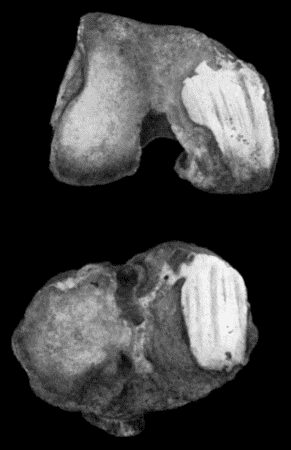
Fig. 158.—Arthritis Deformans of Knee, showing eburnation and grooving of articular surfaces.
(Anatomical Museum, University of Edinburgh.)
Morbid Anatomy.—The commonest type is that in which the articular surfaces undergo degenerative changes. The primary change involves the articular cartilage, which becomes softened and fibrillated and is worn away until the subjacent bone is exposed. If the bone is rarefied, the enlarged cancellous spaces are opened into and an eroded and worm-eaten appearance is brought about; with further use of the joint, the bone is worn away, so that in a ball-and-socket joint like the hip, the head of the femur and the acetabulum are markedly altered in size and shape. More commonly, the bone exposed as a result of disappearance of the cartilage is denser than normal, and under the influence of the movements of the joint, becomes smooth and polished—a change described as eburnation of the articular surfaces (Fig. 158). In hinge-joints such as the knee and elbow, the influence of movement is shown by a series of parallel grooves corresponding to the lines of friction (Fig. 158).
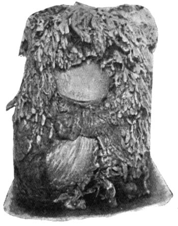
Fig. 159.—Hypertrophied Fringes of Synovial Membrane in Arthritis Deformans of Knee.
(Museum of Royal College of Surgeons, Edinburgh.)
While these degenerative changes are gradually causing destruction of the articular surfaces, reparative and hypertrophic changes are taking place at the periphery. Along the line of the junction between the cartilage and synovial membrane, the proliferation of tissue leads to the formation of nodules or masses of cartilage—ecchondroses—which are subsequently converted into bone (Fig. 157). Gross alterations in the ends of the bone are thus brought about which can be recognised clinically and in skiagrams, and which tend to restrict the normal range of movement. The extension of the ossification into the synovial reflection and capsular ligament adds a collar or “lip” of new bone, known as “lipping” of the articular margins, and also into other ligaments, insertions of tendons and intermuscular septa giving rise to bony outgrowths or osteophytes not unlike those met with in the neuro-arthropathies.
Proliferative changes in the synovial membrane are attended with increased vascularity and thickening of the membrane and an enlargement of its villi and fringes. When the fatty fringes are developed to an exaggerated degree, the condition is described as an arborescent lipoma (Fig. 159). Individual fringes may attain the size of a hazel nut, and the fibro-fatty tissue of which they are composed may be converted into cartilage and bone; such a body may remain attached by a narrow pedicle or stalk, or this may be torn across and the body becomes loose and, unless confined in a recess of the joint, it wanders about and may become impacted between the articular surfaces. These changes in the synovial membrane are often associated with an abundant exudate or hydrops. These degenerative and hypertrophic changes, while usually attended with marked restriction of movement and sometimes by “locking” of the joint, practically never result in ankylosis.
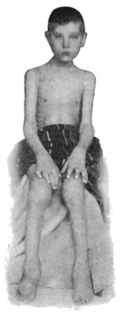
The ankylosing type of chronic arthritis is fortunately much rarer than those described above, and is chiefly met with in the joints of the fingers and toes and in those of the vertebral column. The synovial membrane proliferates, grows over the cartilage, and replaces it, and when two such articular surfaces are in contact they tend to adhere, thus obliterating the joint, cavity, and resulting in fibrous or bony ankylosis. The changes progress slowly and, before they result in ankylosis, various sub-luxations and dislocations may occur with distortion and deformity which, in the case of the fingers, is extremely disabling and unsightly (Fig. 160).
Clinical Features.—It is usually observed that in patients who are still young the tendency is for the disease to advance with considerable rapidity, so that in the course of months it may cause crippling of several joints. The course of the disease as met with in persons past middle life is more chronic; it begins insidiously, and many years may pass before there is pronounced disability. The earliest symptom is stiffness, especially in the morning after rest, which passes off temporarily with use of the limb. As time goes on, the range of movement becomes restricted, and crackings occur. This stage of the disease may be prolonged indefinitely; if it progresses, stiffness becomes more pronounced, certain movements are lost, others develop in abnormal directions, and deformed attitudes add to the disablement. The disease is compatible with long life, but not with any active occupation, hence those of the hospital class who suffer from it tend to accumulate in workhouse infirmaries.
Hydrops is most marked in the knee, and may affect also the adjacent bursæ. As the joint becomes distended with fluid, the ligaments are stretched, the limb becomes weak and unstable, and the patient complains of a feeling of weight, of insecurity, and of tiredness. Pain is occasional and evanescent, and is usually the result of some extra exertion, or exposure to cold and wet. This form of the disease is extremely chronic, and may last for an indefinite number of years. It is to be diagnosed from the other forms of hydrops already considered—the purely traumatic, the pyogenic, gonorrhœal, tuberculous, and syphilitic—and from that associated with Charcot's disease.
Hypertrophied fringes and pedunculated or loose bodies often co-exist with hydrops, and give rise to characteristic clinical features, particularly in the knee. The fringes, especially when they assume the type of the arborescent lipoma, project into the cavity of the joint, filling up its recesses and distending its capsule so that the joint is swollen and slightly flexed. Pain is not a prominent feature, and the patient may walk fairly well. On grasping the joint while it is being actively flexed and extended, the fringes may be felt moving under the fingers. Symptoms from impaction of a loose body are exceptional.
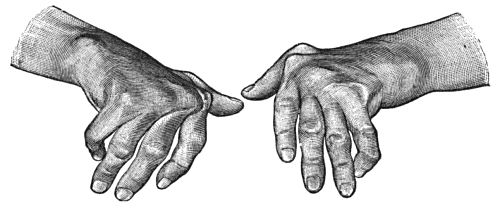
Fig. 160.—Arthritis Deformans of Hands, showing symmetry of lesions, ulnar deviation of fingers, and nodular thickening at inter-phalangeal joints.
The dry form of arthritis deformans, although specially common in the knee, is met with in other joints, either as a mon-articular or poly-articular disease; and it is also met with in the joints of the spine and of the fingers as well as in the temporo-mandibular joint. In the joints of the fingers the disease is remarkably symmetrical, and tends to assume a nodular type (Heberden's nodes) (Fig. 160); in younger subjects it assumes a more painful and progressive fusiform type (Fig. 161). In the larger joints the subjective symptoms usually precede any palpable evidence of disease, the patient complaining of stiffness, crackings, and aching, aggravated by changes in the weather. The roughness due to fibrillation of the articular cartilages causes coarse friction on moving the joint, or, in the knee, on moving the patella on the condyles of the femur. It may be months or even years before the lipping and other hypertrophic changes in the ends of the bones are recognisable, and before the joint assumes the deformed features which the name of the disease suggests.
The capsular ligament, except in hydrops, is the seat of connective-tissue overgrowth, and tends to become contracted and rigid. Intra-articular ligaments, such as the ligamentum teres in the hip, are usually worn away and disappear. The surrounding muscles undergo atrophy, tendons become adherent to their sheaths and may be ossified, and the sheaths of nerves may be involved by the cicatricial changes in the surrounding tissues.
The X-ray appearances of arthritis deformans necessarily vary with the type of the disease and the joint affected; in the joints of the fingers there is a narrowing of the spaces between the articular ends of the bones as a result of absorption of the articular cartilage, and rarefaction of the cancellous tissue in the vicinity of the joints; in the larger joints there is “lipping” of the articular margins, osteophytes, and other evidence of abnormal ossification in and around the joint. Eburnation of the articular surfaces is shown by increase in the density of the shadow of the bone in the areas affected.
Treatment.—Treatment is for the most part limited to the relief of symptoms. On no account should the affected joints be kept at rest by means of splints or other apparatus. Active movements and exercises of all kinds are to be persevered with. When pain is a prominent feature, it may be relieved either by douches of iodine and hot water (tincture of iodine 1 oz. to the quart), or by the application of lint saturated with a lotion made up of chloral hydrate, gr. v, glycerin Ʒj, water ℥j, and covered with oil-silk. Strain and over-use of the joint and sudden changes of temperature are to be avoided. The induction of hyperæmia by means of massage, the elastic bandage, and hot-air baths is often of service. Operative interference is indicated when the disease is of a severe type, when it is mon-articular, and when the general condition of the patient is otherwise favourable. Excision has been practised with success in the hip, knee, elbow, and temporo-mandibular joints. Limitation of movement and locking at the hip-joint when due to new bone round the edge of the acetabulum may be greatly relieved by removal of the bone—a procedure known as cheilotomy. Loose bodies and hypertrophied fringes if causing symptoms may also be removed by operation.
When stiffness and grating on movement are prominent features we have found the injection of from half to one ounce of sterilised white vaseline afford decided relief.
The patient should be nourished well, and there need be no restriction in the diet such as is required in gouty patients, so long as the digestion is not impaired. Benefit is also derived from the administration of cod-liver oil, and of tonics, such as strychnin, arsenic, and iron, and in some cases of iodide of potassium. Luff recommends the administration over long periods of guaiacol carbonate, in cachets beginning with doses of 5–10 grs. and increased to 15–20 grs. thrice daily. A course of treatment at one of the reputed spas—Aix, Bath, Buxton, Gastein, Harrogate, Strathpeffer, Wiesbaden, Wildbad—is often beneficial.
In some cases benefit has followed the prolonged internal administration of liquid paraffin.
On the assumption that the condition is the result of an auto-intoxication from the intestinal tract, saline purges and irrigation of the colon are indicated, and Arbuthnot Lane claims to have brought about improvement by short-circuiting or by resecting the colon.
Residence in a warm and dry climate, with an open-air life, has been known to arrest the disease when other measures have failed to give relief.
The application of radium and the ingestion of radio-active waters have also been recommended.
Hæmophilic or Bleeder's Joint.—This is a rare but characteristic affection met with chiefly in the knee-joint of boys who are the subjects of hæmophilia. After some trivial injury, or even without apparent cause, a hæmorrhage takes place into the joint. The joint is tensely swollen, cannot be completely extended, and is so painful that the patient is obliged to lie up. The temperature is often raised (101° to 102° F.), especially if there are also hæmorrhages elsewhere. The blood in the joint is slowly re-absorbed, and by the end of a fortnight or so, the symptoms completely disappear. As a rule these attacks are repeated; the pain attending them diminishes, but the joint becomes the seat of permanent changes: the synovial membrane is thickened, abnormally vascular, and coloured brown from the deposit of blood pigment; on its surface, and in parts of the articular cartilage, there is a deposit of rust-coloured fibrin; there may be extensive adhesions, and in some cases changes occur like those observed in arthritis deformans with erosion and ulceration of the cartilage and a form of dry caries of the articular surfaces, which may terminate in ankylosis.
As the swelling of the joint is associated with wasting of the muscles, with stiffness, and with flexion, the condition closely resembles tuberculous disease of the synovial membrane. From errors in diagnosis such joints have been operated upon, with disastrous results due to hæmorrhage.
The treatment of a recent hæmorrhage consists in securing absolute rest and applying elastic compression. The introduction of blood-serum (10–15 c.c.) into a vein may assist in arresting the hæmorrhage; anti-diphtheritic serum is that most readily obtainable.
After an interval, measures should be adopted to promote the absorption of blood and to prevent stiffness and flexion; these include massage, movements, and extension with weight and pulley.