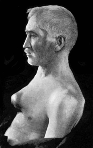
Fig. 74.—Thoracic Aneurysm, threatening to rupture externally, but prevented from doing so by Macewen's needling. The needles were left in for forty-eight hours.
Thoracic Aneurysm.—All varieties of aneurysm occur in the aorta, the fusiform being the most common, although a sacculated aneurysm frequently springs from a fusiform dilatation.
The clinical features depend chiefly on the direction in which the aneurysm enlarges, and are not always well marked even when the sac is of considerable size. They consist in a pulsatile swelling—sometimes in the supra-sternal notch, but usually towards the right side of the sternum—with an increased area of dulness on percussion. With the X-rays a dark shadow is seen corresponding to the sac. Pain is usually a prominent symptom, and is largely referable to the pressure of the aneurysm on the vertebræ or the sternum, causing erosion of these bones. Pressure on the thoracic veins and on the air-passage causes cyanosis and dyspnœa. When the œsophagus is pressed upon, the patient may have difficulty in swallowing. The left recurrent nerve may be stretched or pressed upon as it hooks round the arch of the aorta, and hoarseness of the voice and a characteristic “brassy” cough may result from paralysis of the muscles of the larynx which it supplies. The vagus, the phrenic, and the spinal nerves may also be pressed upon. When the aneurysm is on the transverse part of the arch, the trachea is pulled down with each beat of the heart—a clinical phenomena known as the “tracheal tug.” Aneurysm of the descending aorta may, after eroding the bodies of the vertebræ (Fig. 71) and posterior portions of the ribs, form a swelling in the back to the left of the spine.
Fig. 74.—Thoracic Aneurysm, threatening to rupture externally, but prevented from doing so by Macewen's needling. The needles were left in for forty-eight hours.
Inasmuch as obliteration of the sac and the feeding artery is out of the question, surgical treatment is confined to causing coagulation of the blood in an extension or pouching of the sac, which, making its way through the parietes of the chest, threatens to rupture externally. This may be achieved by Macewen's needles or by the introduction of wire into the sac. We have had cases under observation in which the treatment referred to has been followed by such an amount of improvement that the patient has been able to resume a laborious occupation for one or more years. Christopher Heath found that improvement followed ligation of the left common carotid in aneurysm of the transverse part of the aortic arch.
Abdominal Aneurysm.—Aneurysm is much less frequent in the abdominal than in the thoracic aorta. While any of the large branches in the abdomen may be affected, the most common seats are in the aorta itself, just above the origin of the cœliac artery and at the bifurcation.
The clinical features vary with the site of the aneurysm and with its rapidity and direction of growth. A smooth, rounded swelling, which exhibits expansile pulsation, forms, usually towards the left of the middle line. It may extend upwards under cover of the ribs, downwards towards the pelvis, or backward towards the loin. On palpation a systolic thrill may be detected, but the presence of a murmur is neither constant nor characteristic. Pain is usually present; it may be neuralgic in character, or may simulate renal colic. When the aneurysm presses on the vertebræ and erodes them, the symptoms simulate those of spinal caries, particularly if, as sometimes happens, symptoms of compression paraplegia ensue. In its growth the swelling may press upon and displace the adjacent viscera, and so interfere with their functions.
The diagnosis has to be made from solid or cystic tumours overlying the artery; from a “pulsating aorta”; and from spinal caries; much help is obtained by the use of the X-rays.
The condition usually proves fatal, either by the aneurysm bursting into the peritoneal cavity, or by slow leakage into the retro-peritoneal tissue.
The Moore-Corradi method has been successfully employed, access to the sac having been obtained by opening the abdomen. Ligation of the aorta has so far been unsuccessful, but in one case operated upon by Keen the patient survived forty-eight days.
Innominate aneurysm may be of the fusiform or of the sacculated variety, and is frequently associated with pouching of the aorta. It usually grows upwards and laterally, projecting above the sternum and right clavicle, which may be eroded or displaced (Fig. 75). Symptoms of pressure on the structures in the neck, similar to those produced by aortic aneurysm, occur. The pulses in the right upper extremity and in the right carotid and its branches are diminished and delayed. Pressure on the right brachial plexus causes shooting pain down the arm and muscular paresis on that side. Vaso-motor disturbances and contraction of the pupil on the right side may result from pressure on the sympathetic. Death may take place from rupture, or from pressure on the air-passage.
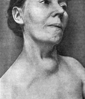
Fig. 75.—Innominate Aneurysm in a woman, æt. 47, eight months after treatment by Moore-Corradi method (cf. Fig. 73).
The available methods of treatment are ligation of the right common carotid and third part of the right subclavian (Wardrop's operation), of which a number of successful cases have been recorded. Those most suitable for ligation are cases in which the aneurysm is circumscribed and globular (Sheen). If ligation is found to be impracticable, the Moore-Corradi method or Macewen's needling may be tried.
Carotid Aneurysms.—Aneurysm of the common carotid is more frequent on the right than on the left side, and is usually situated either at the root of the neck or near the bifurcation. It is the aneurysm most frequently met with in women. From its position the swelling is liable to press on the vagus, recurrent and sympathetic nerves, on the air-passage, and on the œsophagus, giving rise to symptoms referable to such pressure. There may be cerebral symptoms from interference with the blood supply of the brain.
Aneurysm near the origin has to be diagnosed from subclavian, innominate, and aortic aneurysm, and from other swellings—solid or fluid—met with in the neck. It is often difficult to determine with precision the trunk from which an aneurysm at the root of the neck originates, and not infrequently more than one vessel shares in the dilatation. A careful consideration of the position in which the swelling first appeared, of the direction in which it has progressed, of its pressure effects, and of the condition of the pulses beyond, may help in distinguishing between aortic, innominate, carotid, and subclavian aneurysms. Skiagraphy is also of assistance in recognising the vessel involved.
Tumours of the thyreoid, enlarged lymph glands, and fatty and sarcomatous tumours can usually be distinguished from aneurysm by the history of the swelling and by physical examination. Cystic tumours and abscesses in the neck are sometimes more difficult to differentiate on account of the apparently expansile character of the pulsation transmitted to them. The fact that compression of the vessel does not affect the size and tension of these fluid swellings is useful in distinguishing them from aneurysm.
Treatment.—Digital compression of the vessel against the transverse process of the sixth cervical vertebra—the “carotid tubercle”—has been successfully employed in the treatment of aneurysm near the bifurcation. Proximal ligation in the case of high aneurysms, or distal ligation in those situated at the root of the neck, is more certain. Extirpation of the sac is probably the best method of treatment, especially in those of traumatic origin. These operations are attended with considerable risk of hemiplegia from interference with the blood supply of the brain.
The external carotid and the cervical portion of the internal carotid are seldom the primary seat of aneurysm, although they are liable to be implicated by the upward spread of an aneurysm at the bifurcation of the common trunk. In addition to the ordinary signs of aneurysm, the clinical manifestations are chiefly referable to pressure on the pharynx and larynx, and on the hypoglossal nerve. Aneurysm of the internal carotid is of special importance on account of the way in which it bulges into the pharynx in the region of the tonsil, in some cases closely simulating a tonsillar abscess. Cases are on record in which such an aneurysm has been mistaken for an abscess and incised, with disastrous results.
Aneurysmal varix may occur in the neck as a result of stabs or bullet wounds. The communication is usually between the common carotid artery and the internal jugular vein. The resulting interference with the cerebral circulation causes headache, giddiness, and other brain symptoms, and a persistent loud murmur is usually a source of annoyance to the patient and may be sufficient indication for operative treatment.
Intracranial aneurysm involves the internal carotid and its branches, or the basilar artery, and appears to be more frequently associated with syphilis and with valvular disease of the heart than are external aneurysms. It gives rise to symptoms similar to those of other intracranial tumours, and there is sometimes a loud murmur. It usually proves fatal by rupture, and intracranial hæmorrhage. The treatment is to ligate the common carotid or the vertebral artery in the neck, according to the seat of the aneurysm.
Orbital Aneurysm.—The term pulsating exophthalmos is employed to embrace a number of pathological conditions, including aneurysm, in which the chief symptoms are pulsation in the orbit and protrusion of the eyeball. There may be, in addition, congestion and œdema of the eyelids, and a distinct thrill and murmur, which can be controlled by compression of the common carotid in the neck. Varying degrees of ocular paralysis and of interference with vision may also be present.
These symptoms are due, in the majority of cases, to an aneurysmal varix of the internal carotid artery and cavernous sinus, which is often traumatic in origin, being produced either by fracture of the base of the skull or by a punctured wound of the orbit. In other cases they are due to aneurysm of the ophthalmic artery, to thrombosis of the cavernous sinus, and, in rare instances, to cirsoid aneurysm.
If compression of the common carotid is found to arrest the pulsation, ligation of this vessel is indicated.
Subclavian Aneurysm.—Subclavian aneurysm is usually met with in men who follow occupations involving constant use of the shoulder—for example, dock-porters and coal-heavers. It is more common on the right side.
The aneurysm usually springs from the third part of the artery, and appears as a tense, rounded, pulsatile swelling just above the clavicle and to the outer side of the sterno-mastoid muscle. It occasionally extends towards the thorax, where it may become adherent to the pleura. The radial pulse on the same side is small and delayed. Congestion and œdema of the arm, with pain, numbness, and muscular weakness, may result from pressure on the veins and nerves as they pass under the clavicle; and pressure on the phrenic nerve may induce hiccough. The aneurysm is of slow growth, and occasionally undergoes spontaneous cure.
The conditions most likely to be mistaken for it are a soft, rapidly growing sarcoma, and a normal artery raised on a cervical rib.
On account of the relations of the artery and of its branches, treatment is attended with greater difficulty and danger in subclavian than in almost any other form of external aneurysm. The available operative measures are proximal ligation of the innominate, and distal ligation. In some cases it has been found necessary to combine distal ligation with amputation at the shoulder-joint, to prevent the collateral circulation maintaining the flow through the aneurysm. Matas' operation has been successfully performed by Hogarth Pringle.
Axillary Aneurysm.—This is usually met with in the right arm of labouring men and sailors, and not infrequently follows an injury in the region of the shoulder. The vessel may be damaged by the head of a dislocated humerus or in attempts to reduce the dislocation, by the fragments of a fractured bone, or by a stab or cut. Sometimes the vein also is injured and an arterio-venous aneurysm established.
Owing to the laxity of the tissues, it increases rapidly, and it may soon attain a large size, filling up the axilla, and displacing the clavicle upwards. This renders compression of the third part of the subclavian difficult or impossible. It may extend beneath the clavicle into the neck, or, extending inwards may form adhesions to the chest wall, and, after eroding the ribs, to the pleura.
The usual symptoms of aneurysm are present, and the pressure effects on the veins and nerves are similar to those produced by an aneurysm of the subclavian. Intra-thoracic complications, such as pleurisy or pneumonia, are not infrequent when there are adhesions to the chest wall and pleura. Rupture may take place externally, into the shoulder-joint, or into the pleura.
Extirpation of the sac is the operation of choice, but, if this is impracticable, ligation of the third part of the subclavian may be had recourse to.
Brachial aneurysm usually occurs at the bend of the elbow, is of traumatic origin, and is best treated by excision of the sac.
Aneurysmal varix, which was frequently met with in this situation in the days of the barber-surgeons,—usually as a result of the artery having been accidentally wounded while performing venesection of the median basilic vein,—may be treated, according to the amount of discomfort it causes, by a supporting bandage, or by ligation of the artery above and below the point of communication.
Aneurysms of the vessels of the forearm and hand call for no special mention; they are almost invariably traumatic, and are treated by excision of the sac.
Inguinal Aneurysm (Aneurysm of the Iliac and Femoral Arteries).—Aneurysms appearing in the region of Poupart's ligament may have their origin in the external or common iliac arteries or in the upper part of the femoral. On account of the tension of the fascia lata, they tend to spread upwards towards the abdomen, and, to a less extent, downwards into the thigh. Sometimes a constriction occurs across the sac at the level of Poupart's ligament.
The pressure exerted on the nerves and veins of the lower extremity causes pain, congestion, and œdema of the limb. Rupture may take place externally, or into the cellular tissue of the iliac fossa.
These aneurysms have to be diagnosed from pulsating sarcoma growing from the pelvic bones, and from an abscess or a mass of enlarged lymph glands overlying the artery and transmitting its pulsation.
The method of treatment that has met with most success is ligation of the common or external iliac, reached either by reflecting the peritoneum from off the iliac fossa (extra-peritoneal operation), or by going through the peritoneal cavity (trans-peritoneal operation).
Gluteal Aneurysm.—An aneurysm in the buttock may arise from the superior or from the inferior gluteal artery, but by the time it forms a salient swelling it is seldom possible to recognise by external examination in which vessel it takes origin. The special symptoms to which it gives rise are pain down the limb from pressure on the sciatic nerve, and interference with the movements at the hip.
Ligation of the hypogastric (internal iliac) by the trans-peritoneal route is the most satisfactory method of treatment. Extirpation of the sac is difficult and dangerous, especially when the aneurysm has spread into the pelvis.
Femoral Aneurysm.—Aneurysm of the femoral artery beyond the origin of the profunda branch is usually traumatic in origin, and is more common in Scarpa's triangle than in Hunter's canal. Any of the methods already described is available for their treatment—the choice lying between Matas' operation and ligation of the external iliac.
Aneurysm of the profunda femoris is distinguished from that of the main trunk by the fact that the pulses beyond are, in the former, unaffected, and by the normal artery being felt pulsating over or alongside the sac.
In aneurysmal varix, a not infrequent result of a bullet wound or a stab, the communication with the vein may involve the main trunk of the femoral artery. Should operative interference become necessary as a result of progressive increase in size of the tumour, or progressive distension of the veins of the limb, an attempt should be made to separate the vessels concerned and to close the opening in each by suture. If this is impracticable, the artery is tied above and below the communication; gangrene of the limb may supervene, and we have observed a case in which the gangrene extended up to the junction of the middle and lower thirds of the thigh, and in which recovery followed upon amputation of the thigh.
Popliteal Aneurysm.—This is the most common surgical aneurysm, and is not infrequently met with in both limbs. It is generally due to disease of the artery, and repeated slight strains, which are so liable to occur at the knee, play an important part in its formation. In former times it was common in post-boys, from the repeated flexion and extension of the knee in riding.
The aneurysm is usually of the sacculated variety, and may spring from the front or from the back of the vessel. It may exert pressure on the bones and ligaments of the joint, and it has been known to rupture into the articulation. The pain, stiffness, and effusion into the joint which accompany these changes often lead to an erroneous diagnosis of joint disease. The sac may press upon the popliteal artery or vein and their branches, causing congestion and œdema of the leg, and lead to gangrene. Pressure on the tibial and common peroneal nerves gives rise to severe pain, muscular cramp, and weakness of the leg.
The differential diagnosis is to be made from abscess, bursal cyst, enlarged glands, and sarcoma, especially pulsating sarcoma of one of the bones entering into the knee joint.
The choice of operation lies between ligation of the femoral artery in Hunter's canal, and Matas' operation of aneurysmo-arteriorrhaphy. The success which attends the Hunterian operation is evidenced by the fact that Syme performed it thirty-seven times without a single failure. If it fails, the old operation should be considered, but it is a more serious operation, and one which is more liable to be followed by gangrene of the limb. Experience shows that ligation of the vein, or even the removal of a portion of it, is not necessarily followed by gangrene. The risk of gangrene is diminished by a course of digital compression of the femoral artery, before operating on the aneurysm.
Aneurysmal varix is sometimes met with in the region of the popliteal space. It is characterised by the usual symptoms, and is treated by palliative measures, or by ligation of the artery above and below the point of communication.
Aneurysm in the leg and foot is rare. It is almost always traumatic, and is treated by excision of the sac.