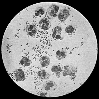
Fig. 2.—Staphylococcus aureus in Pus from case of Osteomyelitis. × 1000 diam. Gram's stain.
From the point of view of the surgeon the most important varieties of micro-organisms are those that cause inflammation and suppuration—the pyogenic bacteria. This group includes a great many species, and these are so widely distributed that they are to be met with under all conditions of everyday life.
The nature of the inflammatory and suppurative processes will be considered in detail later; suffice it here to say that they are brought about by the action of one or other of the organisms that we have now to consider.
It is found that the staphylococci, which cluster into groups, tend to produce localised lesions; while the chain-forms—streptococci—give rise to diffuse, spreading conditions. Many varieties of pyogenic bacteria have now been differentiated, the best known being the staphylococcus aureus, the streptococcus, and the bacillus coli communis.
Staphylococcus Aureus.—This is the commonest organism found in localised inflammatory and suppurative conditions. It varies greatly in its virulence, and is found in such widely different conditions as skin pustules, boils, carbuncles, and some acute inflammations of bone. As seen by the microscope it occurs in grape-like clusters, fission of the individual cells taking place irregularly (Fig. 2). When grown in artificial media, the colonies assume an orange-yellow colour—hence the name aureus. It is of high vitality and resists more prolonged exposure to high temperatures than most non-sporing bacteria. It is capable of lying latent in the tissues for long periods, for example, in the marrow of long bones, and of again becoming active and causing a fresh outbreak of suppuration. This organism is widely distributed: it is found on the skin, in the mouth, and in other situations in the body, and as it is present in the dust of the air and on all objects upon which dust has settled, it is a continual source of infection unless means are taken to exclude it from wounds.
The staphylococcus albus is much less common than the aureus, but has the same properties and characters, save that its growth on artificial media assumes a white colour. It is the common cause of stitch abscesses, the skin being its normal habitat.
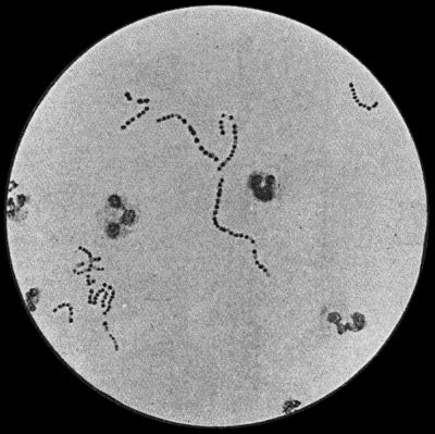
Fig. 3.—Streptococci in Pus from an acute abscess in subcutaneous tissue. × 1000 diam. Gram's stain.
Streptococcus Pyogenes.—This organism also varies greatly in its virulence; in some instances—for example in erysipelas—it causes a sharp attack of acute spreading inflammation, which soon subsides without showing any tendency to end in suppuration; under other conditions it gives rise to a generalised infection which rapidly proves fatal. The streptococcus has less capacity of liquefying the tissues than the staphylococcus, so that pus formation takes place more slowly. At the same time its products are very potent in destroying the tissues in their vicinity, and so interfering with the exudation of leucocytes which would otherwise exercise their protective influence. Streptococci invade the lymph spaces, and are associated with acute spreading conditions such as phlegmonous or erysipelatous inflammations and suppurations, lymphangitis and suppuration in lymph glands, and inflammation of serous and synovial membranes, also with a form of pneumonia which is prone to follow on severe operations in the mouth and throat. Streptococci are also concerned in the production of spreading gangrene and pyæmia.
Division takes place in one axis, so that chains of varying length are formed (Fig. 3). It is less easily cultivated by artificial media than the staphylococcus; it forms a whitish growth.
Bacillus Coli Communis.—This organism, which is a normal inhabitant of the intestinal tract, shows a great tendency to invade any organ or tissue whose vitality is lowered. It is causatively associated with such conditions as peritonitis and peritoneal suppuration resulting from strangulated hernia, appendicitis, or perforation in any part of the alimentary canal. In cystitis, pyelitis, abscess of the kidney, suppuration in the bile-ducts or liver, and in many other abdominal conditions, it plays a most important part. The discharge from wounds infected by this organism has usually a fœtid, or even a fæcal odour, and often contains gases resulting from putrefaction.
It is a small rod-shaped organism with short flagellæ, which render it motile (Fig. 4). It closely resembles the typhoid bacillus, but is distinguished from it by its behaviour in artificial culture media.
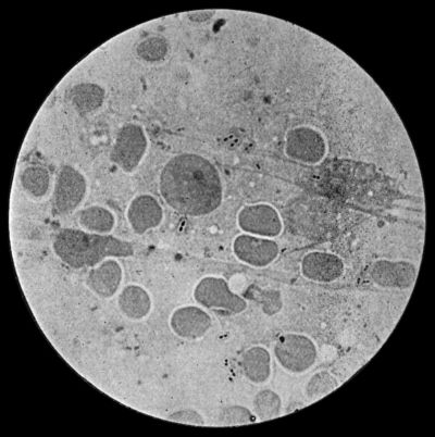
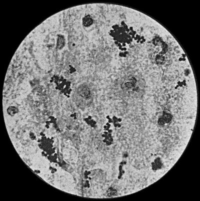
Fig. 5.—Fraenkel's Pneumococci in Pus from Empyema following Pneumonia. × 100 diam. Stained with Muir's capsule stain.
Pneumo-bacteria.—Two forms of organism associated with pneumonia—Fraenkel's pneumococcus (one of the diplococci) (Fig. 5) and Friedländer's pneumo-bacillus (a short rod-shaped form)—are frequently met with in inflammations of the serous and synovial membranes, in suppuration in the liver, and in various other inflammatory and suppurative conditions.
Bacillus Typhosus.—This organism has been found in pure culture in suppurative conditions of bone, of cellular tissue, and of internal organs, especially during convalescence from typhoid fever. Like the staphylococcus, it is capable of lying latent in the tissues for long periods.
Other Pyogenic Bacteria.—It is not necessary to do more than name some of the other organisms that are known to be pyogenic, such as the bacillus pyocyaneus, which is found in green and blue pus, the micrococcus tetragenus, the gonococcus, actinomyces, the glanders bacillus, and the tubercle bacillus. Most of these will receive further mention in connection with the diseases to which they give rise.
Leucocytosis.—Most bacterial diseases, as well as certain other pathological conditions, are associated with an increase in the number of leucocytes in the blood throughout the circulatory system. This condition of the blood, which is known as leucocytosis, is believed to be due to an excessive output and rapid formation of leucocytes by the bone marrow, and it probably has as its object the arrest and destruction of the invading organisms or toxins. To increase the resisting power of the system to pathogenic organisms, an artificial leucocytosis may be induced by subcutaneous injection of a solution of nucleinate of soda (16 minims of a 5 per cent. solution).
The normal number of leucocytes per cubic millimetre varies in different individuals, and in the same individual under different conditions, from 5000 to 10,000: 7500 is a normal average, and anything above 12,000 is considered abnormal. When leucocytosis is present, the number may range from 12,000 to 30,000 or even higher; 40,000 is looked upon as a high degree of leucocytosis. According to Ehrlich, the following may be taken as the standard proportion of the various forms of leucocytes in normal blood: polynuclear neutrophile leucocytes, 70 to 72 per cent.; lymphocytes, 22 to 25 per cent.; eosinophile cells, 2 to 4 per cent.; large mononuclear and transitional leucocytes, 2 to 4 per cent.; mast-cells, 0.5 to 2 per cent.
In estimating the clinical importance of a leucocytosis, it is not sufficient merely to count the aggregate number of leucocytes present. A differential count must be made to determine which variety of cells is in excess. In the majority of surgical affections it is chiefly the granular polymorpho-nuclear neutrophile leucocytes that are in excess (ordinary leucocytosis). In some cases, and particularly in parasitic diseases such as trichiniasis and hydatid disease, the eosinophile leucocytes also show a proportionate increase (eosinophilia). The term lymphocytosis is applied when there is an increase in the number of circulating lymphocytes, as occurs, for example, in lymphatic leucæmia, and in certain cases of syphilis.
Leucocytosis is met with in nearly all acute infective diseases, and in acute pyogenic inflammatory affections, particularly in those attended with suppuration. In exceptionally acute septic conditions the extreme virulence of the toxins may prevent the leucocytes reacting, and leucocytosis may be absent. The absence of leucocytosis in a disease in which it is usually present is therefore to be looked upon as a grave omen, particularly when the general symptoms are severe. In some cases of malignant disease the number of leucocytes is increased to 15,000 or 20,000. A few hours after a severe hæmorrhage also there is usually a leucocytosis of from 15,000 to 30,000, which lasts for three or four days (Lyon). In cases of hæmorrhage the leucocytosis is increased by infusion of fluids into the circulation. After all operations there is at least a transient leucocytosis (post-operative leucocytosis) (F. I. Dawson).
The leucocytosis begins soon after the infection manifests itself—for example, by shivering, rigor, or rise of temperature. The number of leucocytes rises somewhat rapidly, increases while the condition is progressing, and remains high during the febrile period, but there is no constant correspondence between the number of leucocytes and the height of the temperature. The arrest of the inflammation and its resolution are accompanied by a fall in the number of leucocytes, while the occurrence of suppuration is attended with a further increase in their number.
In interpreting the “blood count,” it is to be kept in mind that a physiological leucocytosis occurs within three or four hours of taking a meal, especially one rich in proteins, from 1500 to 2000 being added to the normal number. In this digestion leucocytosis the increase is chiefly in the polynuclear neutrophile leucocytes. Immediately before and after delivery, particularly in primiparæ, there is usually a moderate degree of leucocytosis. If the labour is normal and the puerperium uncomplicated, the number of leucocytes regains the normal in about a week. Lactation has no appreciable effect on the number of leucocytes. In new-born infants the leucocyte count is abnormally high, ranging from 15,000 to 20,000. In children under one year of age, the normal average is from 10,000 to 20,000.
Absence of Leucocytosis—Leucopenia.—In certain infective diseases the number of leucocytes in the circulating blood is abnormally low—3000 or 4000—and this condition is known as leucopenia. It occurs in typhoid fever, especially in the later stages of the disease, in tuberculous lesions unaccompanied by suppuration, in malaria, and in most cases of uncomplicated influenza. The occurrence of leucocytosis in any of these conditions is to be looked upon as an indication that a mixed infection has taken place, and that some suppurative process is present.
The absence of leucocytosis in some cases of virulent septic poisoning has already been referred to.
It will be evident that too much reliance must not be placed upon a single observation, particularly in emergency cases. Whenever possible, a series of observations should be made, the blood being examined about four hours after meals, and about the same hour each day.
The clinical significance of the blood count in individual diseases will be further referred to.
The Iodine or Glycogen Reaction.—The leucocyte count may be supplemented by staining films of the blood with a watery solution of iodine and potassium iodide. In all advancing purulent conditions, in septic poisonings, in pneumonia, and in cancerous growths associated with ulceration, a certain number of the polynuclear leucocytes are stained a brown or reddish-brown colour, due to the action of the iodine on some substance in the cells of the nature of glycogen. This reaction is absent in serous effusions, in unmixed tuberculous infections, in uncomplicated typhoid fever, and in the early stages of cancerous growths.